


We present the case of a 70-year-old male patient with a history of thigh myxoid liposarcoma, sent to our establishment for a CT scan as a part of a follow-up evaluation.
The CT scan revealed multiple metastases in atypical locations, all presenting with similar imaging features. These hypoattenuating formations were identified in various regions: intracardiac (right and left ventricles), mediastinal (posterior mediastinum) (Figure 1), intraperitoneal (splenogastric space and left flank) (Figures 2 and 4), parietal (left abdominal wall) (Figure 3), and in the right scrotum (Figure 5).
Soft tissue sarcomas are rare tumours, comprising 0.7% of all adult malignancies. Liposarcoma is a mesenchymal malignant tumour originating from adipose tissue and can be subclassified into well-differentiated, myxoid, round cell, pleomorphic, and dedifferentiated types. Well-differentiated and myxoid tumours tend to be low-grade with fewer metastases, whereas other histological subtypes present a more invasive profile with a higher metastatic incidence rate [1].
Extremity soft tissue sarcomas typically metastasise to the lungs, but can demonstrate a unique extrapulmonary metastatic pattern. Myxoid liposarcoma, in particular, has a distinct pattern of metastatic spread, favouring extrapulmonary sites such as soft tissue, retroperitoneum, chest and abdominal wall, peritoneal surface, skeleton, and heart [2]. Several studies speculate that the reason behind this specific metastatic spread can be explained by the abundance of fatty tissue in these secondary localisation sites, including subcutaneous tissue, peritoneum, bone marrow, and the epidural space [3].
In this report, we present the case of a lower limb myxoid liposarcoma, a subtype that represents 40% of all liposarcomas of the extremities. The appearance of a myxoid liposarcoma depends on the amount of fat and myxoid tissue. It generally shows a homogeneous low signal on T1 with high-signal foci, a high signal intensity on T2 and variable enhancement based on the cellularity and vascularity of the tumour [3]. In some cases, the tumour appears cystic. Metastases often present a hypodense appearance due to mucinous material, as observed in our patient [4].
The follow-up studies should include a chest, abdominal and pelvic CT, or even a whole-body MRI. Recent studies showed the superiority of this imaging modality in detecting extrapulmonary metastases, especially when it comes to skeletal lesions that are often missed in conventional imaging modalities [5]. However, it remains to be determined whether these findings represent metastases of myxoid liposarcoma or a metachronous disease affecting various locations [6].
The treatment management includes primarily a surgical excision of the tumour with or without adjuvant radiotherapy and chemotherapy. The decision relies entirely on the risk of recurrence. A myxoid liposarcoma is considered to present a poor prognosis and a higher risk of extrapulmonary metastases in case of a large tumour, a high histological grade, and the presence of tumour necrosis [6].
Myxoid liposarcoma metastases
This work is licensed under a Creative Commons Attribution-NonCommercial-ShareAlike 4.0 International License.
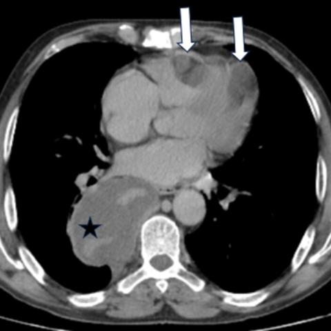


This CT scan demonstrates soft tissue density masses in multiple regions of the chest, abdomen, and pelvis. Specifically:
Overall, these lesions mostly present as low-density with relatively homogeneous or slightly heterogeneous internal signals, showing some mucinous characteristics consistent with previously reported mucoid changes. No obvious signs of bony destruction are noted.
Based on the patient's history (previous diagnosis of thigh myxoid liposarcoma) and the multiple low-density soft tissue lesions shown in this imaging, potential diagnoses include:
Among these, multiple metastases from myxoid liposarcoma are most likely: on one hand, the patient already has a definite pathological diagnosis; on the other hand, the morphology and density observed here align with myxoid soft tissue components. Other diagnoses can be ruled out or supplemented by referencing the patient’s previous medical history, histological features, and imaging findings.
Considering the patient’s age, medical history (thigh myxoid liposarcoma), and the imaging characteristics of multiple low-density soft tissue lesions, the most likely diagnosis is:
Multiple Metastases of Myxoid Liposarcoma
To further confirm the diagnosis, if clinically feasible, biopsy of suspicious lesions or more comprehensive MRI/whole-body imaging evaluation is recommended to delineate their relationship to the primary disease.
For confirmed or highly suspected multiple myxoid liposarcoma metastases, the main treatment principles include:
Rehabilitation and Exercise Prescription Recommendations:
Disclaimer: This report provides a reference analysis and should not be used as the sole basis for final diagnosis or treatment. It is not a substitute for an in-person medical consultation or professional advice. Specific diagnosis and treatment should be made considering the patient’s actual condition and in consultation with relevant specialist departments.
Myxoid liposarcoma metastases