Plantar fascia tear


Clinical History
A 78-year-old male patient suffering from chronic heel pain on weight-bearing experienced a sharp pain with snapping sensation in the plantar aspect of the right heel after prolonged walking. The patient walked with an antalgic gait and physical examination revealed local swelling at the calcaneal attachment of the plantar fascia.
Imaging Findings
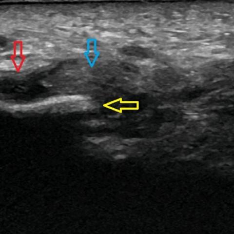
Ultrasound (US) demonstrated a full-thickness tear of the plantar fascia at its calcaneal insertion with a 1,3 cm gap at the site of rupture filled with slightly hypoechoic liquid and tissue reflecting local haemorrhage, oedema and inflammation in the surrounding soft tissues. Retraction and heterogeneity of the ruptured plantar fascia were noted as well as a plantar calcaneal spur.
Discussion
Plantar fascia (PF) disorders result from overuse microtrauma and are aggravated by foot deformities, improper footwear, increased body mass index and weight-bearing physical activities [1]. PF tears are rare and can be divided into acute-on-chronic and acute tears [1,2]. In acute-on chronic cases, the PF tear result as complication of plantar fasciitis (previous steroid injections are a reported risk factor) and usually occur in the proximal fascia [1,3,4]. Acute tears are related to forcible plantar flexion of the foot in competitive athletes and commonly occur distal to calcaneal insertion of the PF[1. Imaging can assist in the diagnosis and rule out other heel pathology. A PF tear should be suspected in the case of sudden heel pain during a weight-bearing activity in a patient with a positive history of plantar fasciitis and/or local corticoid injection. Imaging is of utmost importance for confirming the diagnosis as well as determining the location and severity of the tear (partial or complete).
US findings of plantar fasciitis include increased calibre of the plantar fascia to over 4 mm; loss of reflectivity of the fascia; entheseal new bone formation, including spur formation (a non-specific finding); in severe cases, perifascial collections[1]. The US characteristics of PF tear include complete or partial interruption of the PF and hypoechoic tissue in the region of rupture due to inflammation/haemorrhage. US is superior to MRI in differentiating true fibre tears from oedema [4]. Furthermore, dynamic manoeuvres allow confirmation of a complete tear by demonstrating a gap between the torn parts of the PF[5]. MRI findings of acute PF tear are complete or partial interruption of the low signal of the PF and signal changes at the site of lesion including high signal on fluid-sensitive sequences and intermediate signal on T1-weighted sequences[1]. Plain radiographs add little to the diagnosis of plantar fascia tear but are helpful to rule out concomitant fractures or foot deformities[1,6].
Demonstration of PF tears on imaging is important because treatment differs from plantar fasciitis.
Treatment is based on the severity of the tear and usually involves immobilization and thrombosis prophylaxis, nonsteroidal ant-inflammatory drugs, and physical therapy with a focus on eccentric training [2,6]. Surgical repair is performed only in a few cases of major tears [2,6].
PF tear should be considered in the presence of suggestive clinical history and physical examination. US is a reliable imaging modality to confirm and localize the injury.
Differential Diagnosis List
Final Diagnosis
Complete plantar fascia tear
Liscense
This work is licensed under a Creative Commons Attribution-NonCommercial-ShareAlike 4.0 International License.
Figures

Imaging Findings
Ultrasound imaging shows a noticeable discontinuity of the plantar fascia (PF) near the calcaneal insertion in the right heel, with localized areas of low-echo or anechoic segments suggesting disruption of fascial fibers. Surrounding soft tissues exhibit swelling and mixed echogenicity, possibly related to local inflammation, fluid collection, or hemorrhage. The plantar fascia is thickened (exceeding the normal thickness of about 4 mm), and the echotexture is disordered at the calcaneal attachment. These findings suggest a potential acute tear superimposed on chronic inflammation or overuse injury.
Potential Diagnoses
-
Plantar Fascia Tear
Based on acute heel pain, ultrasound evidence of disrupted fascial fibers at the proximal insertion, and a history of chronic plantar fasciitis, this diagnosis is highly suspected. -
Plantar Fasciitis (Chronic Process)
The lesion may present only with chronic inflammatory thickening without a complete tear; however, the patient has experienced a sudden, tearing-type pain, which, in combination with imaging findings, supports the coexistence of acute and chronic pathology. -
Other Soft Tissue Injury or Bursitis Around the Calcaneus
Typically presents on imaging as soft tissue swelling alone, which does not fully explain the clear fascial fiber disruption seen in this case.
Final Diagnosis
Considering the patient’s age (78 years), history of chronic heel pain, sudden onset of a “tearing” sensation in the plantar area, and ultrasound findings of fascial fiber disruption, the most likely diagnosis is: “Acute Plantar Fascia Tear (Acute on Chronic Plantar Fascia Injury)”.
Treatment Plan & Rehabilitation Program
Based on the extent of the tear, the following management is recommended:
-
Conservative Management
- Relative Immobilization: During the acute phase, reduce weight bearing on the affected foot by using crutches or wearing supportive foot braces (protective shoes) to stabilize foot structures.
- Anti-inflammatory and Pain Relief: A short course of non-steroidal anti-inflammatory drugs (NSAIDs) may help reduce pain and inflammation, but must be considered carefully in light of the patient’s general medical condition.
- Physical Therapy: Once acute pain subsides, gradually initiate ultrasound therapy, alternating cold and warm compresses, massage, and soft tissue release to promote resolution of inflammation and pain relief.
-
Physical Rehabilitation and Exercise Prescription
- Early Stage (Acute Phase, 1–2 weeks): Focus on immobilization, rest, and supportive assistance. Light exercises within a pain-free range, such as toe towel-grasping or heel raises, may be performed in low volume and low repetitions (e.g., 2–3 times per day, each for 10–15 seconds).
- Mid Stage (2–6 weeks): As pain significantly decreases, gradually introduce low-load exercises. Progressively increase eccentric training of the foot arch and posterior lower leg muscles (gastrocnemius, soleus). Increase training frequency by 1–2 sessions per week (each lasting 5–10 minutes), keeping intensity at a low to moderate level. Avoid intense jumping or running.
-
Late Stage (6 weeks and beyond): If the tear has healed sufficiently, broader functional exercises such as walking or light jogging can be introduced. Apply the FITT-VP principles (Frequency, Intensity, Time, Type, Volume, Progression) in an individualized manner, carefully monitoring pain and fatigue:
- Frequency: 3–4 times per week is recommended.
- Intensity: Low to moderate, avoiding any pain exacerbation.
- Time: 20–30 minutes per session, possibly in segments.
- Type: Includes resistance band exercises for ankle muscles, toe-gripping drills, gentle squats, and core stability training.
- Progression: Increase intensity gradually according to rehabilitation progress; avoid sudden excessive loads.
-
Surgical Treatment
Indicated only for severe tears or when sufficient conservative treatment fails to provide relief. The decision depends on tear severity, functional requirements, and preoperative assessment.
Special Precautions: Elderly patients often have osteoporosis, reduced cardiopulmonary function, and other chronic conditions. Exercise prescriptions must be carried out under professional guidance, progressing gradually to prevent falls and related complications.
Disclaimer: This report offers a referential analysis and is not a substitute for an in-person consultation or professional physician’s advice. For a specific treatment plan, please consult an orthopedic or rehabilitation specialist.
Human Doctor Final Diagnosis
Complete plantar fascia tear