Radiological features of Scurvy


Clinical History
A 12-year-old boy presented to the orthopaedic clinic with reduced mobility secondary to bilateral lower limb swelling and severe episodic knee pain. He was noted to have bleeding gums and his examination revealed ecchymosis of both lower limbs. He proceeded to have x-rays and MRI scans of both limbs.
Imaging Findings
MRI scan shows low signal regions on the T1-weighted images which are parallel to the endplates within the distal femoral and proximal tibial metaphysis, which are similarly seen in the proximal fibula [Fig. 2]. Matching areas of increased signal on STIR sequence through the tibial shaft and ankle joint metaphysis are seen[Fig. 3 + Fig. 4]. High STIR signal can also be noted in the distal femur intercondylar notch with low signal on T1-weighted sequences [Fig. 5 + Fig. 6]. There are subperiosteal collections overlying the posterior aspect of distal femurs bilaterally [Fig. 7 + Fig. 8]. Features of myositis within the tibialis anterior muscle and subcutaneous oedema with high signal throughout the medial and lateral gastrocnemius muscles are seen [Fig. 9]. The high STIR sequences are reduced post-treatment, showing radiological improvement [Fig. 10A -11B].
Discussion
Background
Scurvy is a clinical syndrome due to vitamin C deficiency (<11.4 µmol/L). Vitamin C is an essential co-factor required for the hydroxylation of proline and lysine and subsequent collagen synthesis. Its deficiency leads to the production of unstable pro-collagen chains predisposing to weakened bones and increased susceptibility to fractures.
In children, there are a number of risk factors associated with scurvy including malnutrition, malabsorption and neglect. Patients with neurodevelopment or psychiatric conditions are more likely to exhibit food selectivity [1,2]. Conditions that result in iron overload, such as thalassemia also deplete vitamin C causing scurvy [3].
Clinical Perspective
A thorough history including dietary habits and clinical suspicion is imperative to be able to diagnose scurvy. The early signs of scurvy are non-specific (fatigue, irritability and failure to thrive) with the classic symptoms of bleeding gums, ecchymosis, musculoskeletal pain and weakness presenting late. Severe prolonged deficiency can be life-threatening.
Imaging Perspective:
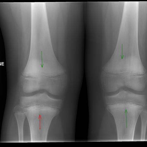
Radiological evidence of scurvy can be seen on plain radiographs of long bones, which are typically symmetrical and bilateral [4,5]:
- Cortical thinning (pencil-point cortex)
- Ground-glass osteoporosis
- Haemarthrosis and periosteal haemorrhages
- Scorbutic rosary: Angular knobbing of the costochondral joints
- Frankel line: Dense zone of provisional metaphyseal calcifications
- Trümmerfeld zone: Radiolucent line that is parallel to Frankel lines
- Wimberger ring: circular calcification surrounding the osteoporotic epiphyseal centre of ossification
- Pelkin spur: Calcification and periosteal elevation beyond the level of metaphysis
- Pelkin fracture: Metaphyseal avulsion fracture
- Corner angle sign: Irregular metaphyseal margins due to small infarctions
- Varus joint deformities and pathological fractures
MRI can detect early changes in scurvy. The commonest finding is diffuse multifocal decreased T1- weighted signal and increased T2-weighted signal within the bone marrow; with the metaphysis being the most affected. [6]
Outcome
The child was diagnosed with scurvy and was commenced on vitamin c replacement. His symptoms fully resolved. Overall, scurvy has an excellent prognosis.
Take-Home Message
Scurvy should be considered as a rare cause for limb and joint pain, particularly in high-risk children. It is important for radiologists to be aware of imaging findings found in scurvy and to appreciate the clinical context.
Differential Diagnosis List
Final Diagnosis
Vitamin C deficiency (Scurvy)
Liscense
This work is licensed under a Creative Commons Attribution-NonCommercial-ShareAlike 4.0 International License.
Figures


I. Radiological Findings
Based on the provided bilateral lower extremity X-ray and knee MRI images, the following main features are noted:
- In the bilateral distal femoral and proximal tibial metaphyseal regions, there are typical abnormalities in density and structure, such as:
- Cortical thinning (resembling a “pencil-thin cortex”) and osteoporosis (“ground-glass-like bone structure”).
- Near the growth plate, linear sclerotic bands (Frankel line) and parallel lucent lines (Trümmerfeld zone) can be observed at the junction between the cortical and cancellous bone.
- The epiphyseal margin may exhibit peripheral sclerosis and ring-like changes (Wimberger ring), suggesting abnormal calcification.
- There may be small local avulsions or a “corner sign” (corner angle sign), along with suspected small fragment loosening (Pelkin fracture) in the proximal tibia.
- Possible signs of bleeding are visible around the joint and beneath the metaphysis, including soft tissue swelling, joint effusion, or a suspected hematoma on X-ray or MRI.
- MRI Findings:
- Multifocal or diffuse bone marrow with decreased T1 signal and increased T2 or STIR signal, especially prominent in the metaphyseal region.
- The soft tissue adjacent to the metaphysis also shows similar high-signal areas, indicating hemorrhage or edema.
II. Potential Diagnosis
Combining the patient’s condition (a 12-year-old male with symmetrical swelling of both lower limbs, recurrent knee pain, gum bleeding, and bruising) and the imaging findings suggestive of malnutrition-related bone changes and hemorrhagic signs, the following differential diagnoses are considered:
- Scurvy:
- Vitamin C deficiency leads to impaired collagen synthesis and predisposes to bone and soft tissue bleeding.
- On imaging, characteristic banded density changes in the metaphyseal region, osteoporosis, and subperiosteal hemorrhages are commonly observed.
- Hemophilia or other coagulation disorders:
- May present with joint hematomas and epiphyseal involvement; imaging often shows recurrent joint bleeding and cartilage destruction.
- Rickets:
- Associated with abnormal calcium and phosphorus metabolism, typically seen as “cup-shaped” deformity of the metaphyses and widened growth plates.
- Leukemic bone infiltration:
- Can cause bone changes and pain, often with diffuse bone marrow infiltration and abnormal complete blood counts.
III. Final Diagnosis
Taking into account the patient’s dietary history, gum bleeding, recurrent bruising, and the aforementioned metaphyseal bone changes and subperiosteal hemorrhage on imaging, the most likely diagnosis is scurvy (Scurvy).
IV. Treatment Plan and Rehabilitation Program
- Treatment Plan:
- Vitamin C Supplementation: Depending on the severity of deficiency, administer oral or intravenous vitamin C following clinical guidelines or specialist advice.
- Supportive Treatment: Pain relief measures and supplementation of other trace elements and vitamins as needed to correct underlying malnutrition.
- If significant joint effusion or subperiosteal bleeding is present, monitor closely or manage further under specialist guidance.
- Rehabilitation Plan and Exercise Prescription:
Once symptoms improve and vitamin C supplementation takes effect, gradually introduce lower limb functional exercises focusing on protecting bones and joints. Refer to the FITT-VP principles:
- Frequency: 3–5 times per week of low-intensity functional and range-of-motion training.
- Intensity: Start with low-intensity exercises to avoid high-impact or excessive load, preventing further injury or fracture.
- Time: Begin with 15–20 minutes per session and gradually increase to 30 minutes based on tolerance.
- Type: Begin with seated or supine active range-of-motion exercises, then gradually transition to low-impact activities such as walking, swimming, or cycling.
- Progression: Increase joint flexion-extension training and light weight-bearing exercises as bone health and symptoms improve, ensuring gradual progression under professional supervision.
- Volume: Start with a lower weekly activity volume and observe the knee joint response. If pain or swelling occurs, adjust the intensity and type of exercise accordingly.
Important note: During the early recovery phase, avoid strenuous running, jumping, or heavy weight-bearing activities to prevent provoking additional bleeding or bone injury.
V. Disclaimer
This report is a reference analysis based on limited information and cannot replace in-person consultations or professional medical advice. All treatment and rehabilitation plans should be tailored to the patient’s actual condition and conducted under the guidance of qualified healthcare professionals.
Human Doctor Final Diagnosis
Vitamin C deficiency (Scurvy)