

A 58-year-old Chinese male with a history of at least 6 years Child-Pugh Class A hepatitis C virus cirrhosis and diabetes mellitus presented with severe pain and swelling in his lower extremities, fever and other septic symptoms (hyperventilation, fast heart rate and confusion) . He indicated vomiting and diarrhoea during a flight from China a few days before his consult, as well as a significant intake of oysters recently.
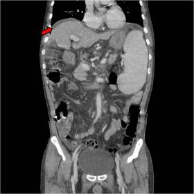
Chest and abdominal X-ray were normal. Contrast-enhanced abdominal CT showed signs of cirrhosis and portal hypertension. However, no infective acute processes were seen (Fig 1). Doppler-US depicted deep vein thrombosis in the lower extremities.
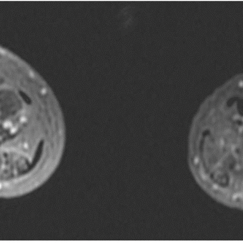
An MRI of the lower extremities was performed. An increase in signal intensity in STIR and T2-weighted images was present in the subcutaneous bilateral fat in thighs and legs, consistent with oedema and cellulitis. Oedema is also seen in multiple muscles in both lower extremities, predominantly around the anterolateral compartment of both legs, suggesting myositis (Fig 2 and 3). Subfascial and intramuscular fluid collections is present, more extensive in the territory of peroneus longus and brevis bilaterally, suggesting abscesses. After the administration of intravenous contrast material, T1 fat-saturated images revealed an enhancement of the muscular fascias and peripheral enhancement of collections (Fig 4 and 5).
Clinical examination and prior imaging exams didn’t reveal any diabetes-associated lower limbs complications.
Background
Vibrio vulnificus is a halophilic gramnegative bacillus found in warmer seawater (temperature>20º), contaminating shellfish and fishes [1], which can cause infections with a wide range of gravity [2].
Clinical perspective
Vibrio vulnificus is usually acquired through the contamination of a previous cutaneous wound or through ingestion. Wound infections can affect immunocompetent or immunocompromised individuals, who can develop from moderate to severe symptoms such as severe cellulitis or necrotizing fasciitis. When a pathogen is acquired through ingestion, most patients develop a limited infection with gastrointestinal symptoms. However, some patients, especially those who are immunocompromised or with diabetes mellitus, end-stage renal disease, rheumatoid arthritis and chronic liver disease, may develop primary septicemia [4,5], with or without gastrointestinal symptoms. Cirrhotic patients are especially vulnerable because V. vulnificus invades the bloodstream by entering the portal system in those with portal hypertension [6, 7]. Septicemia is associated with fever, chills, gastrointestinal symptoms and frequent skin and soft-tissue lesions, primarily in the lower extremities, with bullae, ecchymosis, rash and necrotic ulcer, necrotizing fasciitis and myonecrosis, [8] and finally death. Pneumonia, meningoencephalitis, peritonitis, tuboovarian abscess and pyogenic spondylitis are atypical clinical cases which have been described [9, 10, 11, 12].
Imaging Perspective
MRI is the best imaging technique to identify the severity of soft tissue infection and provide accurate anatomic resolution. Imaging shows non-specific findings, such as subcutaneous and muscular oedema, fluid collections, fascial thickening and enhancement. Intramuscular T1-hyperintensity may represent haemorrhage [13].
These MRI findings in both lower extremities in a septic patient is suspicious of Vibrio vulnificus sepsis, especially with a history a recent fish or shellfish intake. Therefore, MRI helps establishing a proper and early treatment, allowing surgeons to plan abscess draining or debridement if required.
As bowel infection are quite common in those cases, abdominal CT imaging is also a useful tool to support the diagnosis. [14]
Positive blood or wound culture for V. vulnificus confirms the diagnosis [15].
Outcome
V. vulnificus infection is a life-threatening condition. In this context, soft tissue infection, especially necrotizing fasciitis, associates high mortality rates. Properly timed and appropriate antibiotic treatment is extremely important. Nevertheless, early debridement and amputation is sometimes required [3]. In our case, positive blood culture confirmed the diagnosis. Our patient was managed in a conservative manner with intravenous antibiotics, therefore debridement was unnecessary, and lower limbs lesions were recovered.
Take-Home Message / Teaching Points
MRI helps early recognition and treatment, and adequate assessment of soft tissue infection in case surgery is required.
Pyomyositis and cellulitis by Vibrio Vulnificus
This work is licensed under a Creative Commons Attribution-NonCommercial-ShareAlike 4.0 International License.


Based on the provided CT and MRI images, the patient has extensive edema and signal enhancement in the muscles and subcutaneous fascia of both lower limbs. Certain regions show fluid density or signals suggestive of a possible abscess or necrotic lesion. The fascia is thickened and shows enhancement. In some T1-weighted sequences, certain muscle tissues exhibit higher signals, indicating potential hemorrhage or blood stasis. Abdominal CT scans, used to assess possible concurrent intestinal infection, mostly reveal chronic liver changes that may align with a known history of cirrhosis. Overall, these imaging findings are consistent with severe soft tissue infection, including features suggestive of necrotizing fasciitis.
Taking into account the patient’s history of chronic liver disease (HCV-related cirrhosis), immunosuppression from diabetes, recent consumption of raw oysters, and imaging findings indicating severe soft tissue and fascial inflammatory changes in both lower limbs, the final diagnosis is
necrotizing fasciitis and sepsis caused by Vibrio vulnificus.
This diagnosis is also consistent with positive pathogen isolation (V. vulnificus) from blood or wound cultures.
Treatment Strategy
Rehabilitation / Exercise Prescription Advice
This exercise prescription follows the FITT-VP principle (Frequency, Intensity, Time, Type, Volume, and Progression) and should be adjusted according to individual recovery status and overall condition.
Disclaimer: This report is for reference only and does not replace face-to-face diagnosis or professional medical advice. Patients who have any questions or experience worsening symptoms should promptly seek medical attention or consult a healthcare professional.
Pyomyositis and cellulitis by Vibrio Vulnificus