


A one-year-old full-term-born male child, born to non-consanguineous parents, presents with complaints of thoracic deformity. No history of previous surgeries.
The AP chest radiograph (Figure 1) demonstrated the absence of ribs in the right mid-zone, with fusion of multiple adjacent ribs. Fusion of multiple ribs was also noted on the left side. The image showed evidence of dextroscoliosis.
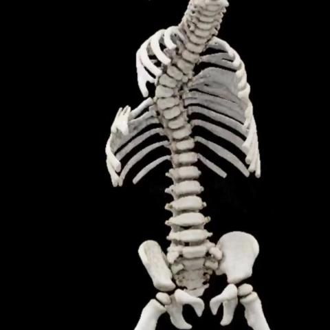
Further evaluation with computed tomography revealed bifid ribs with segmental fusion and widening at multiple levels bilaterally (Videos and Figures 2a, 2b, 2c, 3, 4, and 5). Fusion of posterior vertebral elements was observed at multiple levels, and dextroscoliosis was noted involving the thoracic spine. Posterior vertebral arch defects were identified involving the L5 vertebra and all sacral vertebrae (Figures 2b and 2c).
Mild volume loss of the right hemithorax was noted. The pulmonary parenchyma, bronchial, and vascular markings appeared normal bilaterally.
Spondylocostal dysostosis (SCD), commonly referred to as Jarcho–Levin syndrome, is a rare genetic growth disorder that was first identified by Saul Jarcho and Paul Levin in 1938 [1].
Patients typically exhibit multiple vertebral anomalies across different levels of the spine, such as hemivertebrae, butterfly vertebrae, and fused hypoplastic vertebrae [2]. A common characteristic is the presence of congenital rib malformations, which may be fused, absent, or excessively developed.
Patients may show defects in the neural arches of the vertebrae, potentially resulting in conditions like spina bifida or other neural tube defects. These abnormalities can lead to neurological deficits and complicate the overall clinical presentation.
As a result of these abnormalities, affected individuals have a higher risk of developing thoracic insufficiency syndrome, which can ultimately result in early neonatal death. However, despite these severe complications, around 50% of cases may survive into adulthood [3].
For over fifty years, there has been uncertainty about the differences between two phenotypically similar syndromes that lead to thoracic insufficiency, i.e., spondylocostal dysostosis (SCD), also referred to as Jarcho–Levin syndrome, and spondylothoracic dysostosis (STD), also known as Lavy–Moseley syndrome:
Spondylocostal dysostosis (Jarcho–Levin syndrome)
This work is licensed under a Creative Commons Attribution-NonCommercial-ShareAlike 4.0 International License.


Based on the provided chest X-ray plain films and transverse CT images, the following observations can be made:
Considering the patient’s age (1 year old), chest wall deformities, and imaging features, possible diagnoses include:
Based on the patient’s clinical features (chest wall deformity, absence of notable surgical history), age, and imaging signs of multiple vertebral and rib abnormalities, after excluding other congenital spinal deformities, the most likely diagnosis is:
Spondylocostal Dysostosis (also known as Jarcho–Levin Syndrome).
For confirmation of specific genetic mutations, additional genetic testing is recommended, along with respiratory function assessment and neurological evaluation, to determine the appropriate timing and method of further intervention.
Given the patient’s young age (1 year old), any rehabilitation plan should carefully consider growth and developmental factors.
Disclaimer: This report is for medical reference only and is not intended to replace in-person consultation or professional medical advice. If you have any questions or notice any changes in condition, please seek medical attention promptly and follow the instructions of specialized physicians.
Spondylocostal dysostosis (Jarcho–Levin syndrome)