

Skin lesion (2 centimeters of the largest transverse diameter) clinically suspected to be a melanoma before surgical reject.
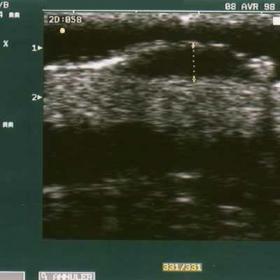
Skin lesion (2 centimeters of the largest transverse diameter) clinically suspected to be a melanoma before surgical reject. A morphological sonographic study was performed to analyse the structural sonographic pattern and to mesure tumor thickness with electronic calipers. The maximal thickness was determined by measuring from the stratum granulosum to the deepest point on the posterior margin. High frequency US scan demonstrates an ovoid hypoechoic tumor with homogeneous echostructure and well-defined lower and lateral margins. The tumor measured: 2.60 mm in thickness (20 MHz) 2.72 mm in thickness (13 MHz) Color Doppler sonography was used to search for intratumoral vessels and completed by a spectral analysis with determination of the resistive index (RI = systolic velocity - diastolic velocity / systolic velocity). The low resistance intra tumor blood flow caracterize the neovascularization. Color Doppler sonography scan reveal a tumor containing intratumor vessels. The arterial velocity spectrum shown below the scan was recorded. The resistive index was low : 0.41
Naevus Current management of melanoma consist in a first surgical resection based on the clinical features followed in about 30% of cases by re-excision according to the Breslow index measured at histological analysis. Surgical margins (according to the french consensus) .must be adapted to the tumor thickness: a marging of at least 1 cm is required for tumors with a Breslow index of 1 mm or less, of 2 cm for those with an index between 1.01 and 4 mm and of 3 cm for those with an index over 4 mm. At histologic analysis, the tumor measured 2 mm in thickness (Breslow index).The overestimation of tumor thickness at US occurrred when a melanoma is developing inside a neavus Also, Color Doppler is able to evaluate angiogenesis and pulsed Doppler characterize the tumor vascularisation of melanoma.
Melanoma measuring 2 mm thickness at histological study. Surgical margins (2 cm) was adapted to the tumor thickness.


Based on the provided ultrasound and color Doppler images, a relatively distinct nodular or raised lesion can be observed in the local area of the skin and subcutaneous tissue. The specific characteristics include:
Considering the patient’s age (51 years), the suspicious clinical lesion (a 2 cm skin lesion), and the abundant blood flow indicated by ultrasound-color Doppler, the main possible diagnoses or differential diagnoses include:
Based on the following factors:
Based on this analysis, the most likely diagnosis is: Malignant Melanoma (Melanoma).
The actual diagnosis should still rely on the patient’s complete medical history, pathological findings, and surgical evaluation. If there is any doubt, further staging assessments such as lymph node ultrasound or other imaging evaluations can be considered to assess possible tumor dissemination.
For postoperative rehabilitation and physical/emotional adjustment, it is important to take into account the surgical wound healing, the location of the lesion, and the overall health status of the patient. The principle is a gradual and individualized approach:
This report is based on the provided basic medical history, imaging, and descriptive information. It offers recommendations for reference only, and cannot replace an in-person consultation or the formal diagnosis and treatment plan of a qualified physician. Patients should follow professional medical advice in conjunction with any clinical assessments and definitive diagnostic conclusions for their treatment and rehabilitation.
Melanoma measuring 2 mm thickness at histological study. Surgical margins (2 cm) was adapted to the tumor thickness.