Calcific Periarthritis of the Finger


Clinical History
A 48 year old female with pain and swelling of the right index finger PIPJ
Imaging Findings
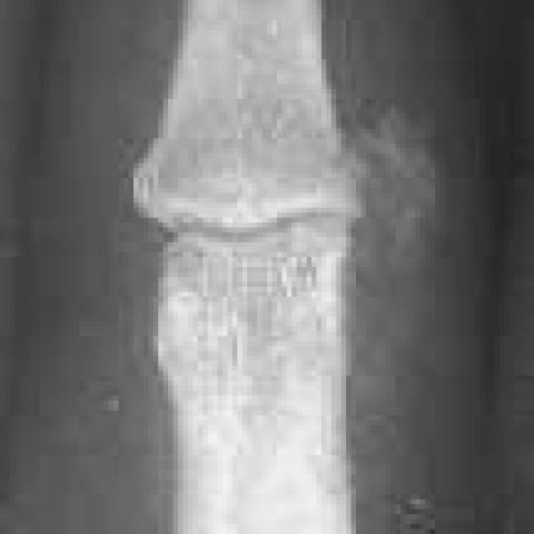
A 48 year old female presented with a two week history of pain and swelling of the right index finger proximal interphalangeal joint (PIPJ). A conventional radiograph of the finger demonstrates an area of amorphous calcification adjacent to the ulnar side of the index finger PIPJ with overlying soft tissue swelling. There is some irregularity of the adjacent cortex of the proximal and middle phalanges (Fig1). A repeat radiograph seven months later demonstrates complete resolution of the calcific deposit. Well defined periarticular erosions with sclerotic margins are seen at the site of the previous cortical irregularity (Fig2).
Discussion
Calcific periarthritis refers to recurrent painful periarticular deposits of calcium in tendons and soft tissues. The deposits are usually monoarticular and the commonest site involved is the shoulder. The hand is less commonly involved, but within the hand, the metacarpophalangeal joints are the commonest sites. The disease may be associated with acute painful episodes or chronic low grade pain and tenderness, but many patients are asymptomatic and the deposits are detected incidentally when radiographed for another reason. The deposits consist of calcium hydroxyapatite crystals. The cause of the crystal deposition is not known but it may be simply secondary to trauma. Radiologically the deposits often appear initially cloud-like, becoming denser and more clearly defined with time. The adjacent bones are usually normal, although contour irregularities, cystic lesions and reactive sclerosis may occasionally occur[1]. The deposits may remain static, enlarge, or reduce in size over time. The diagnosis is important as the symptoms are often misdiagnosed as septic arthritis or gout and inappropriately treated leading to a delay in recovery. The correct treatment consists of immobilisation plus nonsteroidal anti-inflammatory agents. Local steroid injections do not confer any additional benefit. The majority of cases demonstrate resolution of the calcium deposits within a few weeks on follow up radiographs if appropriately treated[2].Several authors have described cases of calcific periarthritis associated with cortical erosions[3, 4]. However, these were all associated with large tendons of the glutei or pectoralis muscles. We have been unable to find any previous descriptions of calcific periarthritis associated with cortical erosions in the digits. Uhthoff and Sarkar[5] describe three distinct stages of calcific periarthritis: precalcific, calcific and postcalcific. The postcalcific stage occurs after the calcific deposit has been phagocytosed and there are said to be no radiographic findings at this stage. This does not concur with our findings in which the cortical erosions persisted after resolution of the calcific deposit. We therefore suggest that calcific periarthritis should be included in the differential diagnosis of periarticular bone erosions.
Differential Diagnosis List
Final Diagnosis
Calcific Periarthritis of the Finger
Liscense
Figures
Conventional radiograph of right index finger

Conventional radiograph of right index finger 7 months later


1. Radiological Findings
Based on the provided X-ray images:
- There is increased soft tissue density around the proximal interphalangeal (PIP) joint of the right index finger, with localized cloudy or dense calcification, confined to the perijoint region.
- The adjacent bony contour is essentially intact, with some minor cortical irregularities or small bony erosions, but no significant widespread bone destruction.
- No marked narrowing of the joint space or severe subchondral sclerosis is observed.
- Soft tissue swelling is primarily localized around the lesion, with no obvious extension to the proximal or distal areas.
2. Possible Diagnoses
Based on the above imaging findings and the patient's background (48-year-old female) with a history of joint pain and localized swelling, the differential diagnoses might include:
- Calcific Periarthritis:
- Commonly presents with cloudy or dense calcifications around the joint, occasionally accompanied by mild cortical changes or erosions.
- More frequently seen in the shoulder joint but can occur in finger joints.
- Often related to repeated microtrauma or degenerative factors. - Gout:
- Typically shows periarticular tophi and bony erosions, often with a “punched-out” appearance, more commonly seen in the first metatarsophalangeal joint but can also involve the fingers.
- In this case, although there are minor cortical erosions, the appearance is not typical for gout, and the clinical history is not fully compatible. - Septic Arthritis:
- Usually presents with marked soft tissue swelling, joint effusion, and bone destruction, along with acute inflammatory symptoms (redness, swelling, heat, and pain).
- This case shows no extensive bone destruction or significant joint space narrowing, making infection less likely.
3. Final Diagnosis
Taking into account the patient's age, symptoms, imaging findings (localized cloudy calcifications, possible mild cortical erosion but overall no significant bone destruction), and comparison with previous reports, the most likely diagnosis is:
Calcific Periarthritis.
After differentiating it from gout and septic arthritis, the clinical and radiological features are most consistent with calcific periarthritis.
4. Treatment Plan and Rehabilitation
4.1 Treatment Strategies
- Conservative Treatment:
- First-line management with nonsteroidal anti-inflammatory drugs (NSAIDs) to relieve pain and inflammation.
- During acute flare-ups, temporary immobilization or splint use can reduce joint load and pain.
- If pain is pronounced, cold compresses or other physical therapy methods may be applied to alleviate local symptoms. - Local Corticosteroid Injection:
- Literature shows that for this type of calcific inflammation, local steroid injections do not necessarily provide significant additional benefit and should be considered cautiously based on the specific condition. - Follow-up and Re-evaluation:
- In most cases, with appropriate conservative management, the calcification can gradually be resorbed or reduced over several weeks.
4.2 Rehabilitation/Exercise Prescription Advice
After acute pain subsides, gradual hand rehabilitation exercises are recommended to prevent joint stiffness and muscle weakness. Following the FITT-VP principle, a progressive program can be arranged:
- Frequency: 1–2 times daily initially, and as pain improves, gradually increase to 3–5 short practice sessions per day.
- Intensity: Avoid causing significant pain; care should be taken to prevent secondary injury or excessive swelling of the joint.
- Time: Begin with 5–10 minutes each session and gradually increase to 15–20 minutes, depending on the patient’s tolerance and recovery.
- Type: Gentle passive joint movements, fist opening and closing exercises, gripping a soft sponge ball, and performing finger range-of-motion exercises after soaking in warm water.
- Progression: Once pain and swelling are substantially reduced, resistance exercises for finger flexion and extension can be introduced cautiously, such as using a light resistance band, provided they do not trigger a recurrence of symptoms.
Patients with other hand or wrist conditions should avoid heavy gripping or prolonged repetitive motions to ensure safety.
Disclaimer
This report is based solely on the provided medical history and imaging data for reference and cannot replace an in-person diagnosis at a regular hospital or a professional physician’s advice. If you have further questions or if symptoms worsen, please seek medical attention promptly.
Human Doctor Final Diagnosis
Calcific Periarthritis of the Finger