Osteosarcoma of the pelvis in an 80 years old woman


Clinical History
An eighty years old woman presented to our hospital complaining of painful swelling of the right inguinal area (firstly noted by the patient one month ago).
Imaging Findings
An eighty years old woman presented to our hospital complaining of painful swelling of the right inguinal area (firstly noted by the patient one month ago). On examination a palpable mass was discovered at the right inguinal area. The conventional x-ray radiograph of the pelvis showed a lytic lesion of the right pubic bone. Ultrasonography showed a mixed echogenity mass. A computed tomography scan with contrast enhancement followed. It showed an expanded erosive lesion with flecks of calcification. Liver, pancreas, spleen, kidneys were normal. The CXR was normal. Retroperitoneal lymph nodes were not found. Laboratory tests showed CA15-3 marker value: 31,2 u/ml (normal 0-28), alkaline phosphatase 157 u/l (normal 30-125). An ultrasound-guided biopsy with 14 gauge cutting needle was performed to establish the final diagnosis.
Discussion
Osteosarcoma is the commonest primary malignant tumour. Typically it appears in adolescent or young adults (10-25 years) slightly predominant in males. It presents usually with localized pain or swelling around the knee. Osteosarcomas are infrequently found in the pelvis and spine. Histologically it is pleomorphic. Two diagnostic features exist: a production of osteoid tissue and presence of alkaline phosphatase within the tumour cells. If cells of cartilage origin are present calcification may be a presenting feature. CT gave us useful information concerning the calcification and the extension of the mass. Biopsy with a cutting needle was performed easily and quickly, as the mass was not very hard. US was useful for avoiding the large inguinal vessels. Our primary diagnosis in this case was chondrosarcoma. Osteosarcoma occurs in older age groups of patients usually associated with Paget’s disease or radiation therapy following treatment of another cancer. In our case there were no signs or history of Paget’s disease, fibrous dysplasia, radiation therapy or existence of osteoblastoma. Furthermore low grade osteosarcoma and parosteal osteosarcoma occur in an older patient population than conventional osteosarcoma.
Differential Diagnosis List
Final Diagnosis
Low grade osteosarcoma (G3/4)
Liscense
Figures
Plain film of the pelvis

Magnified plain film of the pelvis

CT with contrast enhancement of the pelvis

CT with contrast enhancement of the pelvis

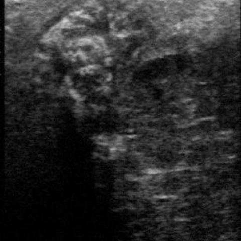
Ultrasound of the pevlis
Imaging Findings
Based on the patient’s X-ray, CT, and ultrasound images, a soft tissue mass is observed in the right inguinal region. Some images suggest possible calcification or osseous signals within the mass. CT indicates that the mass is relatively well-demarcated from the surrounding muscles and superficial soft tissues, although there is a certain degree of local infiltration. During ultrasound examination, the tumor was relatively easy to puncture, indicating that it is not entirely composed of highly ossified or hardened components. The lesion is mainly located in the pelvic area and the inguinal region, with irregular boundaries, suggesting a high suspicion of malignancy involving cartilaginous or bone components.
Potential Diagnoses
- Chondrosarcoma
Reason: Preliminary pathology and imaging suggest cartilage-like or calcification-like changes within the tumor, and chondrosarcomas often present with calcification. Additionally, the location of the lesion (pelvis and surrounding area) is not uncommon for chondrosarcoma. - Osteosarcoma (especially low-grade or parosteal osteosarcoma)
Reason: Osteosarcoma is a common primary malignant bone tumor. While it is most frequently seen in adolescents, it can also occur in older adults, particularly in cases with Paget’s disease or a history of prior radiation. Although there is no evidence of Paget’s disease or prior radiation in this case, the possibility of a low-grade osteosarcoma or parosteal osteosarcoma cannot be ruled out. Findings such as sclerosis, osteoid formation on CT and X-ray, and elevated alkaline phosphatase (if confirmed by laboratory tests) all support this possibility. - Other malignant or metastatic bone tumors
Reason: For example, malignant fibrous histiocytoma (MFH) or metastatic tumors. However, based on the current imaging findings and the patient’s history, these possibilities are relatively lower and require further pathological examination to exclude.
Final Diagnosis
Combining the clinical presentation of an 80-year-old patient with a right inguinal mass, imaging findings showing calcification or osseous tissue, and the discovery of osteoid matrix in the biopsy, the diagnosis currently leans more towards osteosarcoma (low-grade or parosteal). Although chondrosarcoma was initially suspected, if histologic evidence confirms that tumor cells produce osteoid matrix or exhibit distinct malignant bone formation, osteosarcoma would be the more appropriate diagnosis. Ultimately, the final decision will depend on comprehensive pathology and immunohistochemistry reports.
Treatment Plan and Rehabilitation Program
1. Treatment Strategy:
(1) Surgical Treatment: The primary choice is wide or extended resection to ensure a complete surgical margin and reduce the risk of local recurrence.
(2) Chemotherapy or Radiotherapy: Preoperative or postoperative chemotherapy may be considered to reduce residual or metastatic disease, and the specific regimen should be determined by the patient’s overall conditions and pathology results. Radiotherapy generally has lower sensitivity for osteosarcoma, but it can be used as an adjunct if complete resection is infeasible or if local palliation is needed.
(3) Supportive Care: Includes pain management, nutritional support, and bone health optimization according to the patient’s tolerance and systemic condition.
2. Rehabilitation/Exercise Prescription:
Considering the patient’s advanced age and potential surgery and adjuvant therapy, rehabilitation must be individualized, gradual, and follow the FITT-VP (Frequency, Intensity, Time, Type, Progression, Volume) principles:
(1) Early Rehabilitation: During the early phase of treatment or post-surgery, start with simple passive lower limb movements, then gradually progress to bedside sitting and mild resistance training. Frequency: 1-2 times daily, 5-10 minutes each time, with low intensity, ensuring no significant pain or fatigue.
(2) Intermediate Rehabilitation: As the incision heals and pain diminishes, attempt short-distance walking, standing balance exercises, and gentle lower limb strength training with proper protection. Frequency can be increased to 2-3 times daily, 10-15 minutes each, with gradual time extension based on cardiopulmonary capacity.
(3) Late Rehabilitation: If recovery progresses well, gradually increase resistance training intensity with elastic bands, seated or standing leg strengthening exercises, and integrate low-impact aerobic activities (e.g., recumbent cycling). Perform these 3-5 times weekly, 20-30 minutes each session, maintaining joint and soft tissue protection, and following a stepwise approach.
(4) Precautions: Given the patient’s age and relatively fragile bone structure, avoid excessive weight-bearing or intense twisting movements. Pain and vital signs should be closely monitored. Increase load gradually under the guidance of a professional rehabilitation therapist if necessary.
Disclaimer
This report is based solely on current imaging and preliminary pathology information, provided as a reference and without legal authority as the final diagnosis. A definitive diagnosis and treatment strategy should integrate the patient’s complete medical history, laboratory tests, pathological findings, and in-person consultation with specialists. If there are any questions or if symptoms worsen, please promptly consult orthopedics, oncology, or other relevant specialists.
Human Doctor Final Diagnosis
Low grade osteosarcoma (G3/4)