Osteonecrosis in multiple symmetrical lipomatosis


Clinical History
A patient with soft tissue masses in the neck and leg pain.
Imaging Findings
The patient was admitted with an acutely painful left ankle. She gave a year's history of intermittent pain and swelling of the left ankle. The only feature of note from her previous medical history was the excision of two large neck lipomas 20 years earlier. These had since recurred, but further surgery had not been undertaken as she remained asymptomatic. The patient reported that she did not drink alcohol.
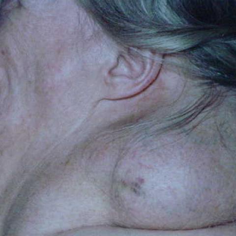
On examination, there were large, bilateral, soft tissue masses extending from the neck to the upper back and shoulders (Fig. 1). The left ankle was red, hot and swollen. Ankle movements were painful.
Haematological and biochemical evaluation was unremarkable apart from mild renal impairment. A clinical diagnosis of cellulitis was made and a rapid recovery ensued after a course of antibiotics.
Plain radiographs of the left leg (Fig. 2) were taken to exclude osteomyelitis. Because of her appearances and the changes noted on plain radiography, MRI was performed for further evaluation. The patient was examined in a Siemens Magnetom 0.2 T open-core system. Images were obtained of both the neck (Fig. 3) and the legs (Fig. 4).
Discussion
Multiple symmetrical lipomatosis (MSL), known also as benign symmetrical lipomatosis, Madelung's disease and Launois-Bensaude disease, is a rare condition first described by Brodie in 1846 and subsequently classified by Madelung and Launois. Its present descriptive name was introduced by Enzi [1].
MSL is rare in the UK and most prevalent in middle-aged Mediterranean males. The dominant clinical feature is painless fat accumulation around the neck, upper back and shoulders. Patients may become symptomatic due to compression of the great vessels and trachea and require surgery. Further manifestations include haematological, metabolic and hepatic disturbances and these are exacerbated by concomitant alcoholism.
Kazumi [2] described a mitochondrial defect leading to alteration of lipolysis in brown fat, the distribution of which is similar to the peculiar position of lipomas in MSL.
MRI [3] helps to determine the extent of the fat accumulation prior to possible surgical resection and also confirms the fatty nature of the subcutaneous masses. A liposarcoma may be identified by an alteration in the otherwise homogeneous MRI signal or by non-suppression in STIR sequences. Cushing's syndrome, which also shows excess fat deposition, is characterised by abnormal biochemisty and osteoporosis.
Osteonecrosis has not been described in MSL previously, however there are multiple associations of altered lipid metabolism and osteonecrosis. Bone infarctions are a recognised complication of hypercortisolism, pancreatitis, diabetes mellitus, alcoholism, and gout as well as haemoglobinopathies, collagen small vessel disease and Caisson disease [4].
Jones [5] examined the relationship of osteonecrosis with different clinical disorders characterised by defective lipid metabolism and suggested a common pathway of continuous or intermittent intraosseus fat emboli leading to focal intravascular coagulation and resulting in osteonecrosis.
Multiple symmetrical lipomatosis with extensive osteonecrosis has not been described previously and the present case might widen the differential diagnosis of bone infarction.
Differential Diagnosis List
Final Diagnosis
Multiple symmetrical lipomatosis
Liscense
Figures
Morphology of MSL
Extensive osteonecrosis in the leg bones


Multiple symmetrical lipomatosis


Osteonecrosis of the proximal tibia


Medical Imaging Analysis Report
I. Imaging Findings
According to the presented imaging data (including the patient’s neck and knee/ankle X-rays and corresponding MRI images), the following main features are observed:
- Cervical Soft Tissue Masses: Multiple symmetrical soft tissue bulges are visible in the posterior neck region and shoulder girdle. MRI reveals that these bulging areas have signals similar to adipose tissue, with a relatively uniform appearance, suggesting a concentrated distribution of a large volume of fat-like components.
- Bone Changes in the Knee and Ankle Joints: X-ray and MRI show local irregular signal changes in the bone, presenting patchy density/signal abnormalities. Areas of bone sclerosis, cystic changes, or features resembling “bone infarction” can be observed; on MRI, lesions similar to bone necrosis (or bone infarction) are irregularly distributed with heterogeneous signals.
- Joint Space: The knee joint space does not appear significantly narrowed, and the cartilage thickness remains acceptable. However, some subchondral bone changes can be noted on X-ray.
II. Potential Diagnoses
Based on the above imaging findings and the patient’s history (69-year-old female with multiple soft tissue masses in the neck and shoulder-back region, as well as leg pain), the following possible diagnoses can be considered:
- Multiple Symmetrical Lipomatosis (MSL):
- This disease, also known as Madelung disease or Launois-Bensaude disease, is a rare benign condition typically characterized by large accumulations of adipose tissue in the neck and shoulder regions.
- The fatty deposits tend to appear symmetrically and are generally painless. On MRI, lipoma-like tissues show signals similar to adipose tissue.
- Liposarcoma:
- If heterogeneous signals or suspicious findings are present within the fatty masses (for instance, on fat suppression sequences), malignant transformation of the fatty component should be considered.
- Osteonecrosis/Bone Infarction:
- Because the patient experiences leg pain and imaging shows bone sclerosis, cystic changes, and heterogeneous signals, osteonecrosis must be taken into account.
- Although osteonecrosis is rarely reported in MSL, given the potential for abnormal lipid metabolism, the occurrence of bone necrosis cannot be entirely ruled out.
- Cushing’s Syndrome and Other Lipid/Endocrine Disorders:
- These conditions can also exhibit abnormal fat distribution. However, patients typically present with clearly elevated cortisol levels and osteoporosis. Current data do not provide clear evidence for this, but they warrant differential consideration.
III. Final Diagnosis
Considering the patient’s clinical symptoms (multiple soft tissue masses in the neck and shoulder-back regions, leg pain), MRI showing symmetrical masses with fat-like signals, and signs in the knee and ankle regions suggestive of osteonecrosis, the most likely best diagnosis is:
Multiple Symmetrical Lipomatosis (MSL) with Concomitant Osteonecrosis (Bone Infarction)
This diagnosis aligns with a rare form of MSL involving abnormal lipid metabolism, leading to possible bone changes and osteonecrosis. If it is necessary to rule out malignant lesions (such as liposarcoma) or other metabolic disorders, further pathological biopsy and additional biochemical/endocrine assessments may be considered.
IV. Treatment Plan and Rehabilitation Program
- Treatment Strategies
- Conservative Treatment: For bone pain and possible pain associated with osteonecrosis, pain medications, physical therapy, and avoidance of excessive weight-bearing can help alleviate symptoms.
- Surgical Intervention: If the accumulation of adipose tissue in the neck region affects the airway, blood vessels, or causes significant cosmetic issues, surgical excision or liposuction may be considered. For confirmed osteonecrotic lesions at risk of collapse or causing severe pain, surgical decompression or repair could be indicated.
- Comprehensive Medical Management: If alcohol abuse or metabolic syndrome is present, targeted interventions should be undertaken. When necessary, medication or other supportive treatments may be recommended to address lipid metabolism, glucose metabolism, and liver function.
- Rehabilitation/Exercise Prescription
- FITT-VP Principle:
- Frequency: Aerobic exercise and moderate-strength training 2-3 times per week, depending on patient tolerance.
- Intensity: Begin with low-intensity activities, such as light walking and simple lower-limb support exercises. Gradually increase intensity based on pain levels and joint stability.
- Time: Start with 10-20 minutes per session and progressively increase to around 30 minutes per session.
- Type: Mild aerobic exercises such as walking or stationary cycling (if knee/ankle issues are not severe), combined with light resistance training (e.g., resistance bands or low-load machines).
- Progression: If no significant discomfort or pain exacerbation occurs, adjust exercise load or duration slightly every 2-4 weeks.
- Volume and Pattern: Exercise can be performed in segments, for example, 10 minutes per session, 2-3 sessions per day. For knee and ankle protection, avoid high-impact activities like running or jumping.
- Individual Adjustments: If the patient has other comorbidities such as reduced cardiopulmonary function or brittle bones, ensure safety when designing the rehabilitation plan. Seek guidance from professional rehabilitation therapists and physicians if needed.
- Key Precautions: Monitor knee and ankle joint pain and range of motion closely. If marked swelling, increased pain, or functional impairment occurs, seek medical attention promptly.
- FITT-VP Principle:
Disclaimer: This report is provided solely as a reference-based analysis on the supplied imaging and medical history and cannot replace in-person consultation or professional medical advice. For specific diagnostic and treatment plans, please consult an appropriate specialist.
Human Doctor Final Diagnosis
Multiple symmetrical lipomatosis