Posterior Glenohumeral Dislocation


Clinical History
Based on the imaging findings the diagnosis of posterior glenohumeral dislocation with fracture of humeral head and neck is made.
Imaging Findings
A patient was admitted to the emergency department after she fell following a bicycle accident. On admission she complained of a painful right shoulder and a painful swelling at the head, the latter corresponding to soft tissue hematoma.
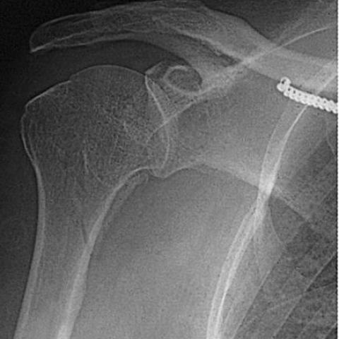
Conventional radiographs of the right shoulder shows on the AP view a spiroid fracture of the surgical neck and proximal diaphysis of the humerus with only minor displacement. Subtle widening of the joint space between the humeral head and anterior lip of the glenoid, but no obvious internal rotation of the humeral head. Posterior oblique view confirms the fracture. There is a small overlap of the humeral head and glenoid.
Scapular Y view shows the posterior orientation of the humeral head in relation to the glenoid. CT scan of the right shoulder includes a section at the level of a middle third of the glenoid which shows posterior dislocation of the humeral head. The humeral head is fixed in this position, as the posterior lip of the glenoid penetrates into a cleft-like defect at the humeral head due to fracture. Section at the level of the lower third of the glenoid demonstrates penetration by the posterior glenoid into the cleft deeper at this level. Clear assessment of the course of the fracture line through the humeral head, ending laterally at the bicipital groove is possible. Section at the level of the surgical neck of the humerus visualizes the fracture lines of the humerus. Displacement is minor. Based on the imaging findings the diagnosis of posterior glenohumeral dislocation with fracture of humeral head and neck is made.
Discussion
Posterior glenohumeral dislocations are uncommon, as they account for only about 2 to 4.3 percent of all shoulder dislocations. It may occur following falls with the arm in adduction, internal rotation and flexion, and following electric shock therapy and seizures. Direct trauma to anterior shoulder may also be the cause, but this is uncommon.
It has been estimated that 40 to 50 percent are overlooked on the initial evaluation, especially on the conventional AP-view radiographs. The following signs are essential in making the diagnosis. On AP views, the humeral head is fixed in internal rotation, the distance between the articular surface of the humeral head and the interior lip of the glenoid may be increased, a vertical line (the 'through' line) may be seen in the medial aspect of the humeral head and indicates the presence of a compression fracture, and finally there may be an associated fracture of the lesser tuberosity. When isolated fractures of the lesser tuberosity are identified, the possibility of associated posterior dislocation should be considered. On 45º posterior oblique view (central X-ray tangentially aligned to the glenoid joint surface), the posterior dislocation manifests as overlap of the humeral head in the glenoid. On scapular Y view (or axillary view), the humeral head projects posteriorly to the glenoid.
CT scan is highly recommended to confirm the presence of the dislocation and to assess the size and course of fracture lines of the impacted fracture of the head of the humerus.
Differential Diagnosis List
Final Diagnosis
Posterior glenohumeral dislocation
Liscense
Figures
Conventional radiographs of the right shoulder


CT scan of the right shoulder


Medical Imaging Analysis Report
1. Imaging Findings
1. X-ray Findings:
- The humeral head is displaced posteriorly relative to the glenoid, showing obvious malalignment between the humeral head and the glenoid fossa, suggesting posterior shoulder dislocation.
- Signs of fracture involving the humeral head and neck are observed, such as an irregular humeral head contour and interrupted local bony trabeculae.
- A fracture line is visible in the humeral neck region, with some bone fragments either separated or overlapping.
- The shoulder joint space appears abnormal, and the humeral head is fixed in an internally rotated position.
- Possible indications of a lesser tubercle (lesser tuberosity) fracture, requiring correlation with CT findings.
2. CT Findings:
- Confirmation of the posterior dislocation of the shoulder joint, with the humeral head clearly displaced posteriorly.
- A compressive fracture is visible on the medial side of the humeral head (posterior dislocations often come with impaction fractures).
- The fracture line between the humeral head and neck is clearly seen, outlining the size and shape of the fracture fragments, some of which may be separated from the main shaft.
- No large discrete bony fragments are observed around the glenoid rim or in the rotator cuff tendon attachment areas, but soft tissue injury should be ruled out through clinical examination.
2. Potential Diagnoses
1. Posterior shoulder dislocation with fractures of the humeral head and neck:
- The most prominent imaging finding is the posterior displacement of the humeral head, along with a visible fracture line.
- Commonly occurs due to electric shock, seizures, or trauma when the arm is in an internally rotated and adducted posture.
- Often associated with fractures of the lesser tubercle or a compressive fracture of the humeral head, aligning with reports in the literature that posterior shoulder dislocations are rare but easily missed.
2. Anterior shoulder dislocation with atypical fractures (differential diagnosis, low probability):
- Certain trauma mechanisms can also cause humeral head fractures, but in anterior dislocation, the humeral head typically shifts anteriorly (or anteroinferiorly).
- Current imaging does not support anterior displacement.
3. Other shoulder-related injuries (soft tissue injuries, partial dislocations, etc.):
- Simple soft tissue lesions or glenohumeral subluxations might show joint space deformations on X-ray or CT, but in this case, there is conclusive evidence of bony dislocation and fracture, making these less likely as the primary diagnosis.
3. Final Diagnosis
Based on the patient’s age, the imaging findings described above, and the clinical history (the typical mechanism leading to posterior dislocation), the most likely diagnosis is:
Humeral Head and Neck Fracture with Posterior Shoulder Dislocation
Further evaluation of the rotator cuff tendons, glenoid labrum, and soft tissues is recommended through physical examination and/or MRI. From the skeletal perspective, however, the posterior dislocation and fracture are clearly established.
4. Treatment Plan and Rehabilitation
1. Treatment Strategies:
- Closed Reduction or Surgical Treatment: For posterior shoulder dislocation, if there are no prominent separated fracture fragments and stability is relatively good, closed reduction under appropriate anesthesia and muscle relaxation may be attempted. If the fracture lines are complex or there is substantial joint surface damage, surgical intervention (e.g., open reduction and internal fixation or arthroplasty) may be necessary.
- Postoperative or Conservative Management: If fracture fragments are unstable, internal fixation is performed to restore stability of the articular surface. Conservative treatment is typically employed for minimal displacement fractures or for patients who cannot tolerate surgery. However, maintaining a reduced position in posterior dislocations can be challenging.
- Pain Management and Anti-inflammatory Therapy: Nonsteroidal anti-inflammatory drugs or other pain relief methods can be used, combined with ice application or physiotherapy to reduce swelling and pain.
2. Rehabilitation and Exercise Prescription (FITT-VP Principle):
- Early Phase (1–2 weeks after surgery or reduction)
· Frequency (F): 2–3 times per day, for about 5–10 minutes each session.
· Intensity (I): Passive or assisted joint movement under professional supervision; movements should be gentle to avoid severe pain.
· Time (T): 5–10 minutes per session, stopping before significant discomfort.
· Type (T): Isometric muscle exercises (static contractions), passive movements of the wrist, elbow, and mild passive movements of the shoulder.
· Progression (P): As pain subsides and the fracture heals, gradually expand the range of motion.
- Intermediate Phase (3–6 weeks after reduction/fixation)
· Frequency (F): 3–5 times per week;
· Intensity (I): Active range-of-motion exercises within a pain-free zone, potentially with resistance bands or light weights;
· Time (T): 15–20 minutes per session, gradually increasing;
· Type (T): Active range-of-motion (AROM) exercises and rotator cuff strengthening;
· Progression (P): As the range of motion improves and the fracture continues healing, intensify muscle strengthening and stability exercises, such as resistance training for the surrounding shoulder muscles.
- Late Phase (6 weeks onward to 3 months)
· Frequency (F): 3–5 times per week;
· Intensity (I): Progressively increase load and range of motion according to rehabilitation evaluations;
· Time (T): 20–30 minutes per session, possibly in divided segments;
· Type (T): Gradual resistance training, proprioception exercises, and functional training (e.g., simulating daily activities);
· Progression (P): Based on follow-up imaging and muscle strength tests, gradually return to daily or sports-related intensity levels.
- Important Notes:
· Ensure the exercise volume is moderate. In case of excessive pain, swelling, or loss of function, seek prompt medical reassessment.
· In patients with osteoporosis, chronic diseases, or compromised cardiopulmonary function, adjust exercise methods and intensity individually.
· Maintain proper shoulder positioning and correct movement patterns, avoiding repeated stress or impact on the shoulder.
5. Disclaimer
This report provides a reference-based analysis derived from available imaging and information. It does not replace in-person medical consultation or a professional physician’s judgment. If further examinations or treatment are required, please seek medical advice and follow the instructions of a specialized physician.
Human Doctor Final Diagnosis
Posterior glenohumeral dislocation