Vertebral sarcoidosis


Clinical History
The patient presented with fever, weight loss and back pain. There was no known history of cancer. X-rays of the spine were normal. Increased vertebral uptake was seen on bone scintigraphy.
Imaging Findings
The patient presented with fever, weight loss and back pain. There was no known history of cancer. X-rays of the spine were normal. Increased vertebral uptake was seen on bone scintigraphy.
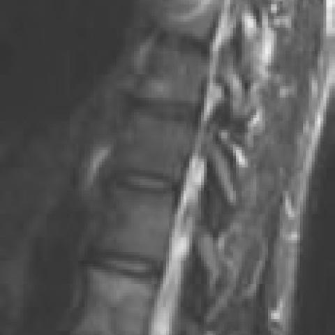
On CT examination multiple thoracic and lumbar vertebral lytic- and sclerotic-appearing lesions were seen. On MRI multiple cervical and thoracolumbar lesions were sdemonstrated (hypointense on T1-weighted images and slightly hyperintense on T2-weighted images) with surrounding bright signal (probably corresponding to oedema) visible around some lesions. No vertebral fracture was visible, and no disk involvement. The epidural space and soft tissues were spared.
Helical chest CT showed mediastinal and bilateral lymph nodes enlargement, thickening of the peribronchovascular bundles, and parenchymal nodules in a peribronchovascular distribution.
Discussion
Sarcoidosis is a multisystem disease characterised by non-caseating granulomas involving essentially the lungs, spleen and lymph nodes. Bone involvement is rare in sarcoidosis, occuring in 1-10% of cases, usually involving the peripheral skeleton. Vertebral involvement has been described in a dozen case reports.
Vertebral lesions usually appear as lytic lesions with sclerotic margins involving the vertebral body and the pedicles (hypointense on T1-weighted images, hyperintense on T2-weighted images). The disk is almost never involved (except in one case report, Kenney et al.). As in our case, a vertebral biopsy is necessary to exclude alternative diagnoses, such as tuberculosis, before initiating steroid therapy. Chest CT is useful to look for associated lymph nodes and parenchymal involvement.
Differential Diagnosis List
Final Diagnosis
Multifocal vertebral sarcoidosis
Liscense
Figures
Thoracolumbar MRI


Total spine CT


Spiral unenhanced chest CT


Imaging Findings
1. From the provided spinal MRI images, multiple abnormal signals can be seen within the vertebral bodies, primarily appearing as low signal on T1-weighted sequences and high signal on T2-weighted sequences. The lesions are confined to the vertebral bodies and pedicles, with no obvious abnormalities seen in the intervertebral discs.
2. CT scans show partial sclerosis of the vertebral bone (with varying degrees) accompanied by relatively well-defined lytic-like changes. The shape of the vertebral bodies is still maintained, and no significant collapse is noted.
3. Chest CT reveals mediastinal and hilar lymphadenopathy as well as mild abnormal findings in parts of the lung parenchyma, suspicious for papular nodules or inflammatory changes.
4. Bone scintigraphy indicates increased uptake in some vertebral segments of the spine, but routine X-ray films do not show typical abnormalities, suggesting early or relatively hidden lesions.
Potential Diagnoses
Based on the clinical presentation (fever, weight loss, and back pain) and imaging findings, possible diagnoses or differential diagnoses include:
1. Spinal Sarcoidosis:
- Characteristics: Non-caseating granulomas often involve hilar lymph nodes and the lungs; spinal involvement is relatively rare.
- Imaging: Lytic lesions may appear within the vertebral bodies, with low signal on T1-weighted MRI and high signal on T2-weighted MRI, typically sparing the intervertebral discs (consistent with this case).
2. Spinal Tuberculosis (Pott’s Disease):
- Characteristics: Common manifestations include fever, weight loss, and other systemic symptoms, as well as vertebral destruction and sequestrum formation. It often involves the intervertebral disc space.
- Imaging: Usually shows lytic destruction of vertebral bodies, narrowing of the intervertebral space, and paravertebral abscess. In this case, the lack of disc involvement is atypical, but this possibility still needs to be ruled out.
3. Metastatic Vertebral Lesions:
- Characteristics: May appear as lytic or mixed lesions. Patients may have a history of primary tumors or unexplained pain.
- Imaging: Multiple vertebral destructions are common. However, in this case, there is no known history of malignancy, and the mediastinal lymph node enlargement suggests another systemic cause, making metastasis less likely.
These diagnoses show some overlap in imaging findings and require correlation with biopsy and additional clinical data for further differentiation.
Final Diagnosis
Considering the patient’s age, clinical presentation (fever, weight loss, back pain), imaging findings (T1 low signal and T2 high signal lesions in the vertebral bodies, with minimal disc involvement), and the exclusion of tuberculosis or other infectious diseases through further biopsy results, the most likely diagnosis is:
Sarcoidosis with Spinal Involvement
Treatment Plan and Rehabilitation
1. Medication: Once the diagnosis of sarcoidosis is established, glucocorticoids (e.g., prednisone) can be administered. The duration depends on disease severity and often lasts for several months or longer. If the patient is unresponsive to steroids or experiences significant side effects, immunosuppressants may be considered.
2. Other Supportive Care: For fever, weight loss, and other systemic symptoms, symptomatic management is recommended, along with adequate nutritional support and appropriate rest. Regular monitoring of lung function and bone status is advised.
3. Rehabilitation and Exercise Prescription (FITT-VP Principle):
- Frequency: 3-5 times per week, depending on patient tolerance and symptom severity.
- Intensity: Start with low-intensity activities (e.g., slow walking, gentle stretching) and gradually increase. In cases of significant back pain, consider non-weight-bearing or low-impact exercises such as swimming or using a stationary bike.
- Time: Each session should last around 20-30 minutes, with the option to break it into shorter segments based on tolerance. Initially, one might begin with 10-minute sessions and repeat multiple times.
- Type: Emphasize flexibility and core stability exercises, including gentle stretching and trunk-strengthening routines (e.g., plank, pelvic bridge), combined with low-intensity aerobic activities.
- Progression: As strength, pain relief, and overall tolerance improve, gradually increase exercise difficulty and duration, avoiding sudden excessive loads.
- Volume & Pattern: When gradually increasing total exercise volume, closely monitor for recurrence of pain or exacerbation of respiratory symptoms, and adjust the training plan accordingly.
Disclaimer
This report provides a reference-based analysis derived from current imaging and clinical information and does not replace an in-person physician consultation or individualized medical advice. If additional clarification is needed, it is recommended to consult a specialist and consider pathology, laboratory tests, and clinical evaluations to reach a final diagnosis and treatment decision.
Human Doctor Final Diagnosis
Multifocal vertebral sarcoidosis