Synovial sarcoma of the acromioclavicular joint


Clinical History
The patient presented with a swelling on the left neck. A gradually increasing mass lesion of approximately 2cm in diameter had been noted 2 months previously and this mass had rapidly increased in size over the previous 2 days. MR imaging was performed.
Imaging Findings
The patient presented with a swelling on the left neck. A gradually increasing mass lesion of approximately 2cm in diameter had been noted 2 months previously and this mass had rapidly increased in size over the previous 20 days. On physical examination, there was a firm mass lesion in the left supraclavicular region extending to the left shoulder and neck. The mass was fixed to surrounding tissues. There was no lesion in the right supraclavicular region. Laboratory results were within the normal limits.
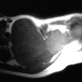
Magnetic resonance (MR) imaging was performed. On T1-weighted MR images, there was an isointense mass lesion relative to surrounding muscle tissue in the supraclavicular region extending to the neck. There were also hypointense signals within the lesion (Fig. 1a). The lesion showed prominent contrast enhancement after contrast medium injection and the hypointense component became more prominent (Fig. 1b). The solid component of the lesion displayed isointense signals relative to surrounding muscle tissue on fast spin-echo T2-weighted MR images (Fig. 1c). The necrotic component within the lesion showed increased signals. MR angiography showed a large feeding artery originating from the left vertebral artery (Fig. 1d).
Discussion
Synovial sarcoma is a rare tumour that constitutes approximately 10% of all soft tissue sarcomas. It is most common in the 2nd and 4th decades of life but may be seen in all age groups (1). The name synovial sarcoma originates from the similarity of the cells with synovial cells which line the surface of joints. Synovial sarcoma is mostly located in tendons and bursae in close proximity to joints. It generally involves the lower extremities, but may be seen in the upper extremities, head and neck region, the wall of the abdomen, and the ankle. The location in the supraclavicular region is relatively rare. In this location it generally originates from the acromioclavicular joint, and it is relatively difficult to differentiate it from lymph node enlargement, which is most commonly seen in that region due to metastatis. The young age of the patient and the absence of findings of primary tumour should alert the physician. Synovial sarcoma of the head and neck region seems to carry a better prognosis than synovial sarcoma of the extremities.
On plain radiographies, soft tissue densities with well-defined contours can be detected in 67% of cases located near joints. Calcifications can be seen in 30% of cases. Bone destruction is detected in 20% of cases because of a pressure effect of the lesion on neighboring bone. The lesion shows increased density compared with muscle on CT scans. CT is better than MR imaging in demonstrating cortical bone involvement and calcifications. Sometimes the lesions show a cystic or haemorrhagic component. Although it is not possible to make a specific diagnosis with MR imaging, some MR imaging features have been described in the literature. Synovial sarcoma can be seen as a heterogeneous multiloculated mass lesion with internal septations. The contour of the lesion is generally well-defined and may have a capsule formation (1-4). The lesions show isointense signals relative to muscle tissue on T1-weighted MR images and hypo- or hyperintense signals on T2-weighted MR images. Recent studies about the dynamic Gd-enhanced imaging in synovial sarcomas showed enhancement of syonovial sarcomas within 7 s after arterial enhancement. This finding was found to be a consistent sign mostly encountered in this tumor (5).
Differential Diagnosis List
Final Diagnosis
Synovial sarcoma of the left acromioclavicular joint
Liscense
Figures
MR imaging appearance of synovial sarcoma


Imaging Findings
The patient is a 28-year-old female with a progressively enlarging mass palpable in the left side of the neck. Based on the provided MRI images, there is a soft tissue lesion located in the left neck near the supraclavicular fossa region, characterized by:
- Shape: The lesion has relatively well-defined margins, appearing fairly regular or slightly lobulated. Some areas show septations or small cystic components.
- Signal Characteristics: On T1-weighted images, the lesion’s signal is similar to or slightly lower than muscle. On T2-weighted images, it appears as a heterogeneous high signal with uneven internal signals, suggesting possible cystic or hemorrhagic components.
- Enhancement Pattern: Dynamic contrast-enhanced scans show early enhancement, indicating a relatively rich blood supply.
- Local bone destruction or compression is not clearly evident (if CT reveals cortical changes, it would suggest involvement of adjacent bone surfaces). However, assessment here mainly relies on MRI.
Potential Diagnoses
Considering the patient’s young age, rapid growth of the mass, and the MRI findings of well-defined margins and enhancement features, the following differential diagnoses are possible:
- Synovial Sarcoma: Commonly occurs in adolescents and middle-aged adults, can arise in the head and neck region or around joints in the extremities. Imaging often shows a well-demarcated soft tissue mass with internal septations and prominent contrast enhancement.
- Metastatic Lymph Node or Lymphoma: Enlarged cervical lymph nodes are common. However, most do not show marked septation or cystic changes. If metastatic, the presence of a primary lesion should be considered.
- Other Malignant Soft Tissue Tumors (e.g., Malignant Peripheral Nerve Sheath Tumor, Fibrosarcoma): Some can present as mixed signals with high vascularity. Clinical and imaging features may overlap with synovial sarcoma, so histopathological examination is required for definitive classification.
Final Diagnosis
Based on the clinical history (young patient, palpable neck mass, rapid growth), imaging findings (well-defined margins, septations, multiple signal components, rapid enhancement), and the fact that such tumors are rare in the head and neck region but documented in the literature, the most likely diagnosis is: Synovial Sarcoma.
Since imaging alone cannot completely rule out other soft tissue tumors, a definitive diagnosis requires histopathological and immunohistochemical examination. If necessary, further evaluation through biopsy or surgical resection with pathological assessment is recommended.
Treatment Plan and Rehabilitation
Treatment Strategy
For synovial sarcoma and other malignant soft tissue tumors, a multidisciplinary approach is advised:
- Surgical Treatment: Generally remains the primary treatment. The goal is complete resection of the tumor with clear surgical margins. The extent of surgery depends on tumor staging and location, aiming to preserve function while removing the lesion in its entirety.
- Radiotherapy and Chemotherapy: For some high-grade soft tissue sarcomas, adjuvant radiotherapy can be considered before or after surgery. Chemotherapy regimens are determined by histological subtype and tumor staging.
- Targeted or Immunotherapy: Depending on molecular pathology and recent research advances, certain patients may benefit from targeted therapies or immunotherapy. This requires genetic testing and expert evaluation.
- Follow-up and Review: Regular imaging (MRI or CT) and relevant blood tests to promptly detect any recurrence or metastasis.
Rehabilitation and Exercise Prescription
Treatment via surgery or radiotherapy/chemotherapy can affect physical function, including local soft tissue and joint mobility, as well as overall endurance and muscle strength. Rehabilitation should be individualized, with reference to the following key points:
- F (Frequency): 3–5 rehabilitation sessions per week, gradually increasing to daily low-intensity activities as tolerated.
- I (Intensity): Start with low intensity (RPE indicating slightly increased breathing rate but still able to carry on a conversation), then progress to moderate intensity.
- T (Time): Each session of about 15–30 minutes, which can be divided into smaller intervals. Session duration can be extended as fitness improves.
- T (Type): Considering neck and shoulder function and overall condition, include stretching exercises for the neck and shoulder region, low-intensity aerobic exercises (such as walking on a treadmill or cycling). Avoid high-impact activities or excessive traction on the affected area.
- V (Volume): Gradually increase total exercise volume within a safe range, taking care not to overexert.
- P (Progression): Reassess pain levels, joint mobility, and fatigue every 2–4 weeks, and appropriately increase training intensity or introduce new activities, such as light strength training or aquatic exercises.
During rehabilitation, avoid heavy lifting, intense impact, or overstretching of the affected neck area. If persistent pain, swelling, or other discomfort occurs, seek medical evaluation promptly.
Disclaimer: This report is based solely on the provided imaging data and limited clinical information for initial assessment. It does not replace an in-person medical consultation or professional medical advice. Specific treatment and rehabilitation plans should be made under the guidance of a specialist.
Human Doctor Final Diagnosis
Synovial sarcoma of the left acromioclavicular joint