Fibrous dysplasia of the skull


Clinical History
Bony swelling on the head.
Imaging Findings
The patient presented with a slowly enlarging bony swelling on the head, present for many years. There was no pain or neurological symptoms.
Discussion
Fibrous dysplasia, also known as Jaffe-Lichtenstein disease, is a developmental anomaly of the skeleton of unknown cause. It occurs in monostotic and polyostotic forms, seen in 70-80% and 20-30% of cases respectively. Of these 2-3% are associated with endocrinopathies, typically the McCune-Albright syndrome. The skull and facial bones are affected in 10-25% of monostotic cases and 50% of polyostotic cases. The common sites are the frontal, sphenoid, maxillary and ethmoid bones, with the temporal and occipital being less common. The lesions are usually asymptomatic but may present with deformity or neurological complications, such as optic nerve compression.
On plain radiographs there may be single or multiple lesions, which are predominantly sclerotic but may be lucent. When expansion occurs it tends to be outwards with bulging of the outer skull table, which remains intact. Computed tomography is useful to show the extent of bone involvement, as well as the extracortical involvement. The main differential diagnosis is Paget's disease where the features of facial bone involvement and prominence of the external occipital protruberance are less common and the skull tables may be destroyed. Hypervascularity of fibrous dysplasia of the skull is an unusual association which may present with recurrent haemorrhage.
Differential Diagnosis List
Final Diagnosis
Fibrous dysplasia
Liscense
Figures
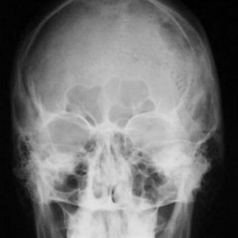
Plain film of skull

CT of skull


Post-operative skull X-rays


1. Imaging Findings
Based on the imaging provided by the patient (cranial X-ray and CT), the following features can be observed:
• Localized thickening of the skull, mainly involving the left side (or corresponding region), showing relatively uniform dense or sclerotic changes;
• The outer table of the skull frequently exhibits outward bulging; however, its continuity remains visible, with no obvious signs of destruction;
• On CT images, the bone in the lesion area presents a “ground-glass” or sclerotic appearance, and the distinction between cortical and cancellous bone in the medullary cavity is not particularly clear;
• No significant soft tissue mass or intracranial parenchymal involvement is observed, although the localized bone expansion is pronounced, warranting vigilance for possible neural canal compression.
2. Potential Diagnoses
Combining the patient's age, clinical history, and imaging findings, the following diagnoses or differential diagnoses are considered:
- Fibrous Dysplasia (Jaffe-Lichtenstein Disease): Commonly presented as monostotic or polyostotic lesions, often involving craniofacial bones. On imaging, characteristic findings include a “ground-glass” appearance, bone densification, and expansile changes. In most cases, the outer table remains intact.
- Paget’s Disease (Osteitis Deformans): Can also present with skull thickening, often accompanied by irregular destruction and new bone formation in the inner and outer skull tables, typically described as “cotton wool-like” imaging changes. If facial bones are involved or there is a notably enlarged external occipital protuberance, and laboratory measurements (such as elevated alkaline phosphatase) are consistent, Paget’s disease becomes more likely.
In summary, fibrous dysplasia and Paget’s disease are common differential diagnoses for skull thickening and expansion. Given the localized lesion and the relatively intact outer table in this case, Paget’s disease is less likely.
3. Final Diagnosis
Taking into account the patient’s age (26-year-old male), symptoms (bony palpable mass on the head), imaging characteristics (localized expansion, noticeable “ground-glass” sclerotic area, preservation of the outer table), and the typical predilection for craniofacial involvement in fibrous dysplasia, the most likely diagnosis in this case is:
Fibrous Dysplasia (Fibrous Dysplasia).
If clinical uncertainty persists, further imaging follow-up (to observe progression) or, when appropriate, a histopathological biopsy may be considered to confirm the diagnosis.
4. Treatment Plan and Rehabilitation
4.1 Treatment Strategy
• Observation and Follow-up: For patients with mild symptoms or no significant functional impairment, regular follow-up can be considered to assess changes in lesion size, shape, and any signs of neural compression.
• Surgical Treatment: If the lesion causes a notable cosmetic deformity, affects the optic canal or other cranial nerve pathways, or creates psychological distress, surgical intervention (such as lesion curettage/decompression, reconstructive repair) can be considered.
• Medication: Currently, there is no specific medication that can reverse fibrous dysplasia. However, for patients experiencing worsening bone pain or significant bone destruction, bisphosphonates may be used to alleviate symptoms under the guidance of a specialist.
4.2 Rehabilitation and Exercise Prescription
Because fibrous dysplasia can weaken the affected bone, exercise should be introduced progressively with appropriate safety measures. According to the FITT-VP principle (Frequency, Intensity, Time, Type, Progression, Individualization), the following suggestions are made:
- Frequency: Engage in moderate exercise 3–5 times per week, such as walking, light jogging, simple fitness exercises, or swimming.
- Intensity: Initially select low to moderate intensity (for instance, maintaining a heart rate at 50%–60% of the maximum) and avoid high-impact or vigorous contact sports to reduce the risk of fractures or injury.
- Time: Aim for 20–30 minutes of activity per session, progressively increasing duration based on the patient’s physical condition and bone health.
- Type: Focus on low-impact aerobic activities (e.g., brisk walking, cycling, swimming) combined with mild resistance exercises (e.g., elastic band workouts, light resistance training). Under professional supervision, incorporate exercises to strengthen core stability and flexibility.
- Progression: As pain and fatigue improve, the duration and intensity of exercise may be gradually increased, closely monitoring any skeletal discomfort or neurological symptoms.
- Individualization: If the patient has other health issues (e.g., reduced cardiopulmonary function), a more cautious program should be devised in consultation with physicians and rehabilitation specialists, with periodic imaging if necessary.
During all activities, avoid impacts or external pressure to the head and facial region, preventing uneven bone stress that may lead to occult fractures or nerve compression. In case of severe headache, localized tenderness, or worsening neurological symptoms, seek medical attention promptly.
5. Disclaimer
This report is based on the patient’s provided medical history and imaging data for analysis and is intended solely as a reference for medical information. It does not substitute for an in-person consultation or professional medical advice. If you have any questions or develop new clinical symptoms, please promptly consult a specialist for further evaluation.
Human Doctor Final Diagnosis
Fibrous dysplasia