Bone involvement in secondary syphilis: Proliferative periostitis and destructive osteitis.


Clinical History
A 45-year-old male presented to the emergency department with a two-month history of pain and swelling of the left thumb. He also mentioned both tibias, right elbow and left ankle pain and swelling, febricula and weight-loss. The patient had no recent high-risk sexual behaviour but referred a history of treated syphilis 7 years ago.
Imaging Findings
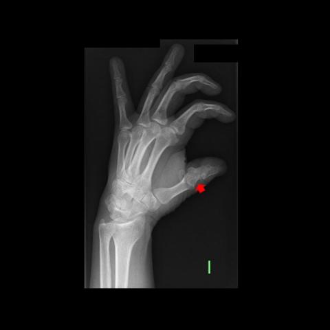
Anteroposterior and lateral radiographs (Fig. 1a, b) of the left thumb showed a permeative lytic lesion in the proximal phalanx associated with pathological fracture.
Anteroposterior and lateral radiographs (Fig. 2a, b, and c) of both tibias revealed subcortical lytic lesions in the anterior bone cortex and interrupted periosteal reaction.
Computed tomography (CT) (Fig. 3a, b, and c) and fat-suppressed proton density MRI (Fig. 4) of both tibias revealed subcortical lytic lesions with a small soft tissue component. An aggressive-appearing periosteal reaction surrounds the lesions. MRI demonstrated no bone marrow involvement. The diagnosis of bone syphilis lesions was suggested.
Skeletal scintigraphy (Fig. 5) revealed increased uptake in both tibias, areas of the hands, the ankles and the feet. The report suggested lung cancer metastasis.
Positron emission tomography scan (PET-CT) was subsequently performed which did not provide further information (Fig 6a). No primary tumour was identified.
After antibiotic treatment follow-up, PET-CT (Fig. 6b) showed significantly decreased uptake in both tibias and left ankle. Oblique radiograph of the left hand (Fig 7) revealed fracture healing of the proximal phalanx of the thumb.
Discussion
Patient outcome and follow-up
A quantitative rapid plasma reagin (RPR) test was performed with a titer of 1/64 and a fluorescent treponemal antibody absorption test showed a positive result. Serology could be consistent with a new infection, inadequate treatment before infection or expected serologic findings post-treatment.
Pathological examination of tibia lesions showed unspecific inflammatory cells and no evidence of malignancy. Immunohistochemical stain demonstrated no treponema.
The patient was treated empirically with intravenous penicillin for two weeks and on-demand analgesics. No additional treatment was performed. Symptomatic relief occurred rapidly after the start of the antibiotic. Follow-up PET-CT showed markedly decreased uptake of the bone lesions. The antibody titer significantly diminished (1/16) and the finger fracture healed 11 months later.
Background
Syphilis is a sexually transmitted disease caused by the spirochete Treponema pallidum. Musculoskeletal manifestations can be associated with congenital, secondary, and tertiary syphilis. However, although bone involvement is a common finding in congenital syphilis, it is rare in secondary syphilis [1].
Reynolds and Wasserman review the largest series of early acquired syphilis in 1942. They found only 15 cases with bone involvement of 10000 cases of early acquired syphilis (0.15%) [2].
A recent English- language literature review from 1964 to 2013 found 36 cases of secondary syphilis with bone involvement [3]. In this review, the diagnosis of early syphilis was suspected based on mucocutaneous findings in 76% of patients. In the remaining 24% of the patients there was no history of syphilitic mucocutaneous lesions, and high-titer nontreponemal serologic tests were the only evidence of early syphilis, as in our case. The most frequent skeletal involvement was in the long bones of the limbs and the skull [3].
Bone pain was the most common complaint. Examination may reveal tenderness over the involved bones and local oedema.
Bone involvement is usually in the form of proliferative periosteal reaction and, less frequently, destructive osteitis and osteomyelitis. Lytic lesions, mainly in the long bones, and aggressive periosteal reaction surrounding the lesion are typical findings [1,2,3,4]. Scintigraphy may show polyostotic uptake that can mimic malignancy [5]. Imaging is important both in the diagnosis and in the assessment of response to treatment. Pathologic examination usually reveals non-specific inflammatory infiltration. Identification of spirochetes in the bone lesion with dark-field microscope, silver stain, immuoperoxidase stain, or polymerase chain reaction is uncommon, and only 5 cases have been reported in the English literature [3].
The availability of imaging modalities such as CT, MRI, and bone scintigraphy has led to easier diagnosis and more frequent reporting of osseous syphilis than in the past [4]. Imaging can detect silent or even asymptomatic bone lesions.
Syphilis is a treatable disease with antibiotics [6]. Untreated, it may lead to severe disease [5].
It appears that the incidence of bony involvement in early syphilis may be higher than previously thought [3,4,5]. It is important to keep in mind this diagnosis, since the imaging appearance may mimic other diseases such as malignancy.
Written informed patient consent for publication has been obtained.
Differential Diagnosis List
Final Diagnosis
Bone involvement in secondary syphilis: proliferative periostitis and destructive osteitis
Liscense
This work is licensed under a Creative Commons Attribution-NonCommercial-ShareAlike 4.0 International License.
Figures


Imaging Findings
Based on the provided X-ray, CT, and MRI images, the following findings are observed:
- Left Thumb: Localized abnormal bone density in the proximal or middle phalanx of the left thumb, showing focal bone destruction and periosteal reaction. Mild to moderate swelling of the surrounding soft tissue is also noted.
- Bilateral Tibias: The tibial shaft shows significant periosteal proliferation (“onion-skin” or layered appearance) and localized reduction or irregular changes in bone density. CT reveals thickening of the tibial surface and cortex, whereas MRI shows corresponding bone marrow edema and mild soft tissue involvement.
- Whole-body Bone Scan (SPECT/PET, etc.): Increased radiotracer uptake in multiple sites, including both tibias, the left ankle, the right elbow, and the left thumb, indicating multifocal active bone lesions.
The overall imaging presentation shows multiple bones and joints involved, with both proliferative periosteal reactions and localized destructive changes. Considering the clinical context, possible causes include infection or neoplastic disease, among others.
Potential Diagnoses
- Infectious Bone Disease (Syphilitic Osteitis, Tuberculosis, Other Bacterial or Fungal Infections): The patient has a history of syphilis, and current serology shows an RPR titer of 1:64 and a positive FTA-ABS, suggesting active syphilis. Imaging may show periosteal proliferation and focal bone destruction, which are consistent with syphilitic osteitis. Other infections such as tuberculosis or fungal infections could also cause bone destruction and periosteal proliferation, but correlation with clinical history and laboratory findings is required.
- Malignant Tumors (Primary or Metastatic): For instance, osteosarcoma or metastatic cancer can present with bone destruction and aggressive periosteal reactions. However, such cases often show more aggressive imaging features (e.g., Codman’s triangle or large soft tissue masses). Multi-site symptoms and systemic changes should be correlated with tumor markers and pathology to rule out malignancies.
- Chronic Osteomyelitis or Non-specific Inflammation: May manifest as bone destruction, periosteal proliferation, and sclerosis. Typically, there is clear evidence of pyogenic bacteria or other pathogens, or long-standing clinical signs such as persistent sinus tracts.
Final Diagnosis
Considering the patient’s age (45), clinical presentation (multiple bone pain and swelling, low-grade fever, weight loss), history of syphilis treatment, current serological findings (RPR 1:64, FTA-ABS positive), multifocal bone lesions on imaging, and rapid response to antibiotic therapy, the most likely diagnosis is:
Syphilitic Osteitis (Early Syphilitic Infection Involving Bones)
Negative results for syphilis spirochetes on pathological examination do not exclude the diagnosis, as it can be challenging to detect the pathogen in bone lesions. Ultimately, clinical correlation and improvement on imaging help confirm the diagnosis.
Treatment Plan and Rehabilitation
Based on the principles of treating early syphilitic bone involvement, the first-line treatment is penicillin antibiotics. Common clinical practice includes:
- Medication: Continuous intravenous penicillin for at least 2 weeks, with possible extension according to the patient’s condition. If the patient is allergic to penicillin, second-line antibiotics should be used, and serological markers must be closely monitored.
- Symptomatic Management: Non-steroidal anti-inflammatory drugs (NSAIDs) or other analgesics can be used to relieve pain and inflammation. If there is no significant instability such as a fracture, immobilization or bracing can be considered as appropriate.
During bone healing and overall recovery, rehabilitation training is crucial. The FITT-VP principle (Frequency, Intensity, Time, Type, Volume/Progression) can guide the rehabilitation process:
- Acute Phase (Early Treatment)
- Frequency: 2–3 times per week, balancing rest and therapy
- Intensity: Low-intensity isometric exercises to avoid excessive strain on the bones
- Time: About 10–15 minutes per session, adjusted for tolerance
- Type: Light activities for the affected regions and adjacent joints, such as grip exercises, ankle mobility, and core stability training
- Subacute Phase (After Marked Symptom Improvement)
- Frequency: 3–4 times per week
- Intensity: Gradually transition to moderate intensity, e.g., light-resistance exercises (elastic bands or small dumbbells)
- Time: 20–30 minutes per session
- Type: Comprehensive exercises to enhance lower and upper limb muscle strength and joint flexibility; focus on joint range of motion and muscle endurance
- Rehabilitation Phase (Later Consolidation Stage)
- Frequency: 4–5 times per week, avoiding excessive overload
- Intensity: Increase resistance or training duration gradually if no pain or adverse local reactions occur
- Time: At least 30 minutes per session, extended as needed
- Type: Progressive return to normal exercise routines (e.g., light jogging, swimming, low-impact aerobic exercises) to maintain and improve overall cardiovascular fitness and muscle strength
During recovery, it is essential to monitor for pain or swelling. If any abnormalities occur, prompt re-evaluation and adjustment of the training program are necessary. For patients with fragile bones or other chronic conditions, individualized exercise plans should be developed to avoid overly aggressive weight-bearing exercises.
Disclaimer: This report is based solely on the provided medical history, imaging data, and related materials. It cannot replace an in-person consultation or professional guidance and treatment by a qualified physician. Specific treatment should be determined by a specialist’s comprehensive evaluation of the patient’s clinical situation.
Human Doctor Final Diagnosis
Bone involvement in secondary syphilis: proliferative periostitis and destructive osteitis