


Painful swollen left elbow with incomplete extension following judo injury.
The patient presented to A&E with a painful left elbow sustained whilst engaging in judo. The arm was swollen and there was incomplete extension. Plain films of the elbow were taken.
At birth both the ends of the forearm and humerus that make up the elbow are cartilaginous. During childhood, from approximately 6 months to 12 years, the six elbow ossification centres form.
The exact age at which these ossification centres appear in not as important as the order in which they appear. The order of ossification is given by the acronym CRITOL.
C Capittelum
R Radial head
I Internal (medial) epicondyle
T Trochlea
O Olecranon
L Lateral (external) epicondyle
The principle of the sequence is that a latter ossification centre should not be seen without the former being seen. Unfortunately the sequence is not always followed, but the trochlea invariable ossifies after the internal epicondyle. Thus, if the trochlea is seen, the internal epicondyle must be seen elsewhere on the epicondyle.
Paradoxically, the grossly displaced internal epicondyle may be more difficult to see that a minimally displaced epicondyle. This is because the grossly displaced epicondyle tends to overlie the joint and may be misinterpreted as another centre.
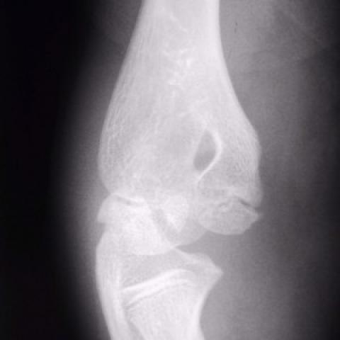
The films show an elbow effusion as demonstrated by a posterior fat sign, which occurs as the medial epicondyle is intra-capsular. The trochlea is seen to be present, but the internal epicondyle is not normally located. It overlies the joint space on the lateral view, and can be seen projected over the capitellum on the frontal view.
Early identification of an avusled medial epicondyle is required as the fragment is almost always displaced inferiorly, but may also be displaced anteriorly or posteriorly as well and may enter the joint space, commonly between the trochlea and the coronoid process of the ulna, and cause entrapment.
Avulsed elbow medial epicondyle


Based on the provided X-rays:
Based on the above radiological features and clinical symptoms (pain, swelling, limited mobility after a sports injury), the possible diagnoses include:
Considering the patient’s age, clinical presentation (traumatic injury during judo training), imaging features (local swelling, fat pad sign, displaced medial epicondyle fragment), and the timing of epiphyseal appearance, the most likely diagnosis is:
Medial Epicondyle Avulsion Fracture of the Humerus.
Principles of rehabilitation: gradual progression and individualization.
Throughout the rehabilitation process, regular follow-ups are necessary to adjust the exercise program based on recovery progress. If significant pain or discomfort occurs, stop the relevant activities and consult a physician or rehabilitation therapist.
FITT-VP Principle Example:
This report is solely a reference analysis based on the provided imaging and history. It cannot replace an in-person consultation or professional medical advice. If you have any questions or if symptoms worsen, please seek immediate medical attention or consult a specialist.
Avulsed elbow medial epicondyle