


The patient presented with a painful deformed right forearm following a 4ft fall from a play gym onto a toy. Examination revealed a tender right forearm, reduced elbow movements but no neurovascular deficit.
The patient presented with a painful deformed right forearm. He had fallen 4 feet from a play gym with his right forearm landing awkwardly on a toy. Examination revealed a distressed boy with an obviously deformed right forearm, which was tender to palpation. The elbow was held in 60 degrees of flexion and the patient was reluctant to allow flexion or extension of the elbow. There was no neuro-vascular deficit and he did not sustain any other injury.
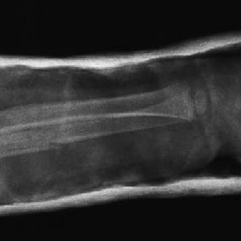
Radiographs of the right forearm were taken showing a segmental fracture of the ulna with an anterior dislocation of the radial head (Fig. 1).
A closed reduction was achieved with longitudinal traction of the forearm and flexion of the elbow with posterior thumb pressure on the radial head (Fig. 2). The arm was immobilised in a posterior above-elbow slab for the first week then converted to a full above-elbow and immobilised for 5 weeks.
Fractures of the radius and/or ulna shafts are relatively common, accounting for 5-10% of children's fractures. In 1814, Giovanni Monteggia described two cases of fracture of the proximal third of the ulna associated with anterior dislocation of the radial head. Bado later gave the eponym of Monteggia lesion. There are 4 types of Monteggia fracture-dislocations. ·Type 1 Anterior dislocation of the radial head with fracture of the ulna at any point and anterior angulation. ·Type 2 Posterior dislocation of the radial head associated with fracture of the ulna and posterior angulation. ·Type 3 Lateral dislocation of the radial head associated with fracture of the ulna and lateral angulation. ·Type 4 A fracture of the upper third of the radius and ulnar shafts with anterior dislocation of the head of the radius. The fracture of the ulnar shaft is angulated anteriorly. In type 1 direct force to the ulna or a hyper-pronation indirect force due to a fall on the outstretched hand is the mechanism of fracture. Monteggia equivalent fractures are variants of type I, one of which describes a single/segmental fracture of the ulna with an associated fracture of the radial neck and dislocation of the shaft of the radius. This case reports a low velocity injury to a 3 years old child, which resulted in a closed segmental fracture of the ulna with an anterior dislocation of the intact radial head. This type of Monteggia equivalent fracture has not been described previously. Fractures of the radius and ulna shafts can almost always be successfully treated with closed reduction and cast immobilisation. Indications for operative treatment include arterial injury, compartment syndrome, open fractures, irreducible fractures, and failure to maintain an adequate reduction.
A segmental fracture of the right ulna with an anterior dislocation of the radial head


Based on the provided right forearm X-ray, the following main features are observed:
1. There is a clear segmental fracture of the ulna with abnormal alignment of the fracture segments.
2. The proximal radius appears intact, but the radial head is dislocated anteriorly (toward the front).
3. Soft tissue swelling around the fracture site and increased radiolucency suggest soft tissue injury or edema.
4. No obvious fracture of the elbow joint itself was noted, but the abnormal relationship of the humeroradial joint indicates the radial head has moved out of its normal articulation.
5. There is no significant radiographic sign of neurovascular injury, although minor injuries cannot be completely ruled out; clinical observation is necessary.
Based on the mechanism of injury, clinical presentation, and radiographic features, the following diagnoses or differential diagnoses might be considered:
1. Classic Monteggia Fracture (Type I)
Given that the patient has a proximal or metaphyseal ulna fracture along with an anterior dislocation of the radial head, this is consistent with the basic features of a Type I Monteggia fracture (anterior dislocation).
2. Monteggia Equivalent Injury
The patient presents with a segmental fracture of the ulna, and the radial head, albeit intact, is dislocated. This aligns with a rare “Monteggia equivalent” variant. Some literature also refers to such presentations as Monteggia equivalent injuries, requiring attention to differentiate and accurately name.
3. Simple Radius and Ulna Shaft Fractures
If the radial head dislocation were overlooked, this might simply be considered a forearm fracture (radius and ulna). However, the X-ray confirms a radial head dislocation, making this possibility less likely.
Based on the patient’s age, mechanism of injury, imaging findings, and the presence of a segmental ulna fracture combined with an anterior radial head dislocation, the most likely diagnosis is “Monteggia Equivalent Injury, approaching a Type I Monteggia variant.”
1. Treatment Principles
• Closed Reduction and Cast Immobilization: Most pediatric forearm fractures can be managed with closed reduction under manipulation, ensuring anatomical alignment of the ulna fracture and the reduction of the radial head dislocation, followed by long-arm casting. If the reduction is satisfactory and stability can be maintained, continued follow-up observation is recommended.
• Surgical Intervention: Surgery may be necessary under the following circumstances:
2. Rehabilitation and Exercise Prescription
Rehabilitation focuses on the gradual recovery of limb function, including forearm rotation, elbow flexion and extension, and wrist function. A step-by-step approach following the FITT-VP principle is recommended:
This report is a reference medical analysis based on the provided data and cannot substitute for an in-person consultation or professional medical evaluation. If the condition changes or any emergency arises, please seek immediate medical attention.
A segmental fracture of the right ulna with an anterior dislocation of the radial head