Solitary sacral plasmocytoma


Clinical History
The patient presented with a five-year history of low back pain which had become very severe and constant over the past ten days. The pain was mechanical in nature and he was unable to sleep due to the pain. There was associated radicular pain in the distribution of the first left sacral nerve root, but no symptoms relating to the bowel or bladder. Physical examination revealed mild tenderness in the upper aspect of the sacrum. Neurological and digital rectal examinations were normal.
Imaging Findings
The patient presented with a five-year history of low back pain which had become very severe and constant over the past ten days. The pain was mechanical in nature and he was unable to sleep due to the pain. There was associated radicular pain in the distribution of the first left sacral nerve root, but no symptoms relating to the bowel or bladder. Physical examination revealed mild tenderness in the upper aspect of the sacrum. Neurological examination was normal and so was digital rectal examination. His haematological and biochemical blood parameters and urinalysis were within normal limits. The patient was admitted for pain relief and further investigations.
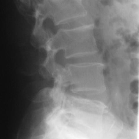
Plain radiographs of the lumbo-sacral spine (Fig. 1) did not reveal any significant abnormality. A CT scan revealed a lesion in the body of the first sacral vertebra extending into the left lateral mass with encroachment of the spinal canal and compression of the cauda equina and bilateral first sacral nerve roots (Fig. 2). A CT-guided biopsy of the lesion was performed (Fig. 3). The patient's serum electrophoresis was normal and his urine was negative for Bence Jones proteins. There were no hot spots on his bone scan (Fig. 4), but the lower sacrum was not visible as he failed to empty the bladder. Five days later the histopathology of the lesion came back as plasma cell neoplasia. A skeletal survey did not reveal any abnormality other than degenerative changes in the lumbar spine. Bone marrow aspiration cytology from the iliac crest was also within normal limits. This being a solitary plasmacytoma in the sacrum with no bone marrow involvement, he was treated with radiotherapy to the lesion.
Discussion
Sacral tumours often present as low back pain with no characteristic pattern or time course. These tumours can be large at presentation as a large volume of tumour can be accommodated within the pelvis (1). Pre-sacral tumours can sometimes be palpated on rectal examination. Encroachment into the spinal canal or the neural foramina can cause cauda equina compression. This is manifested as varying degrees of neurological deficit in the lower limbs with bowel or bladder dysfunction.
Solitary bone plasmacytomas are rare plasma cell proliferative disorders. Most often they involve the bones of the axial skeleton. In the spine, the vertebral body or the posterior elements can be involved.
Plain radiographs of sacral lesions may look normal in spite of extensive involvement of the bone. CT images demonstrate myelomatous mass with the same radiodensity as the adjacent musculature.
The diagnosis is based on histological confirmation of monoclonal plasma cell infiltration of a single disease site and on the exclusion of systemic myeloma by aspiration of marrow from an uninvolved bone (usually the iliac crest). Normal marrow contains about 3% plasma cells. If the aspirate contains a significantly greater proportion of undifferentiated or differentiated plasma cells, the disease is considered disseminated. But there exists a tendency in not accepting the 'solitary bone plasmocytoma' as a true isolated tumor mass. Although no or only very few tumor cells (smoldering myeloma) are visible in the bone marrow aspirate, all these 'solitary' tumors will generalize with time.
For solitary spinal lesions, the treatment of choice is localised radiotherapy (2). Chordoma is the most common primary bone tumour of the sacrum. Metastatic disease, giant cell tumours, lymphomas and myelomas are the other frequent neoplasms of the sacrum (3). Enostosis, osteoid osteoma, osteoblastoma, aneurysmal bone cyst, osteochondroma, chondrosarcoma, Ewing sarcoma, primitive neuroectodermal tumour, and osteosarcoma should also be considered in the differential diagnosis (3).
Differential Diagnosis List
Final Diagnosis
Solitary sacral plasmocytoma
Liscense
Figures
Plain radiographs of the lumbosacral spine


CT images at the level of S1


CT-guided biopsy

Bone scan

Medical Analysis Report
I. Radiological Findings
Based on the provided lumbosacral X-ray and CT images, the following features were observed:
- Mild bone destruction or abnormal texture in the upper sacrum. On some plain radiographs, only irregular radiolucent areas with unclear margins can be identified.
- CT images show locally reduced density lesions, with density comparable to or slightly higher than the surrounding soft tissues, consistent with typical lytic lesions.
- No significant canal stenosis was noted near the sacral canal, but attention should be paid to possible infiltration of the lesion into neural foramina or surrounding soft tissues.
- No signs of acute fracture or significant vertebral collapse were observed in the overall lumbar series.
II. Possible Diagnoses
Considering the patient is a middle-aged male with chronic lumbosacral pain that has recently worsened, and radiographic findings show a lytic lesion in the sacrum, the following possible diagnoses should be considered:
- Solitary Bone Plasmacytoma: Multiple myeloma often affects the axial skeleton. A solitary bone lesion can manifest as a lytic lesion, with CT density similar to or slightly higher than surrounding soft tissue. Further evaluation with bone marrow aspiration and pathological confirmation is required.
- Chordoma: Commonly arising in the sacrococcygeal region, it is one of the most frequently seen primary bone tumors of the sacrum. Imaging typically demonstrates lytic destruction and may be associated with a soft-tissue mass.
- Metastatic Tumor: If the patient has a history of other tumors, metastases should be considered. In the absence of an identified primary site, metastatic disease remains part of the differential diagnosis.
- Giant Cell Tumor: Seen in young to middle-aged adults, frequently located in the sacrum. X-ray findings often show a soap-bubble or lytic pattern of bone destruction.
- Lymphoma or Other Rare Bone Tumors: When bone is involved, lymphoma can present with lytic lesions and should be included in differential considerations.
Further clinical correlation with laboratory tests (hematology, biochemistry, tumor markers) and pathological results is necessary to confirm or rule out these possibilities.
III. Final Diagnosis
According to the histopathological findings, there is monoclonal plasma cell infiltration. Meanwhile, bone marrow aspiration from “unaffected” sites did not show significant tumor cell proliferation. Combining the clinical symptoms, radiological appearance, and laboratory evidence, the final diagnosis is:
Solitary Bone Plasmacytoma of the Sacrum.
If further clarification is needed, a biopsy of the lesion or an extended bone marrow biopsy may be performed to exclude diffuse multiple myeloma and other focal lesions in the body.
IV. Treatment Plan and Rehabilitation
The standard treatment for solitary bone plasmacytoma usually involves local radiotherapy, with surgical decompression or lesion resection when necessary. If the disease progresses or evolves into multiple myeloma, systemic therapy with chemotherapy may be considered.
1. Treatment Strategies
- Local Radiotherapy: Use radical or palliative radiotherapy on the lesion site. Dosage depends on tumor size and pathological confirmation.
- Surgical Intervention: For intractable pain, neurological symptoms, or pathological instability, consider surgical decompression with internal fixation or lesion resection.
- Medication: Under the guidance of a physician or hematologist, corticosteroids, immunomodulators, or other medications may be prescribed based on disease status and overall patient condition.
- Regular Follow-up: Regular MRI, CT, or blood tests are recommended to monitor disease progression and multiple myeloma-related indicators (e.g., serum protein electrophoresis).
2. Rehabilitation and Exercise Guidelines (FITT-VP Principle)
Rehabilitation should follow a principle of gradually progressing from low to moderate intensity with an individualized approach. The following suggestions may be considered:
- Type: Select activities that place less stress on the lumbosacral region, such as short walks, using an elliptical machine, swimming, or gentle stretching.
- Frequency: Start with three sessions of light exercise per week, increasing to four or five times per week once the condition is stable.
- Intensity: Begin at low-to-moderate intensity, with perceived exertion at a “light to moderate” level. The RPE (Rate of Perceived Exertion) can be maintained around 11–13.
- Time: Each session can start at 15–20 minutes. Depending on tolerance and recovery, increase by 5–10 minutes every one to two weeks, gradually reaching over 30 minutes.
- Variation: As lumbosacral pain improves, gradually add core exercises such as prone back extensions or gentle yoga movements, avoiding excessive twisting or high-impact activities.
- Progression: Once pain is controlled, gradually increase the intensity and duration of exercise. Carefully monitor for any recurrence of pain or worsening neurological symptoms, and consult a rehabilitation specialist or physician as needed.
Additionally, pay attention to bone quality. If there is osteoporosis or a high risk of pathological fracture, safety measures should be strictly observed to prevent impact or falls.
Disclaimer:
This report is based on the patient’s provided history and imaging data for reference only. It does not substitute an in-person consultation or professional medical advice. If you have further questions or if the patient’s condition changes, please consult a specialist promptly.
Human Doctor Final Diagnosis
Solitary sacral plasmocytoma