Osseous Kaposi's sarcoma


Clinical History
HIV-positive patient with an osteolytic lesion.
Imaging Findings
This patient, who had been HIV-positive for 8 years and suffered from cutaneous Kaposi's syndrome for 6 years, underwent chemotherapy (ABV-HCG) and radiotherapy (3500rads). The lesions were located on the chest, left arm, left thigh and left calf. In May 2000, the cutaneous lesions worsened, with redness and painful swelling of the patient's left arm in the region of his elbow. X-rays showed a large osteolytic lesion of the ulna, and so regional radiotherapy and systematic chemotherapy were begun.
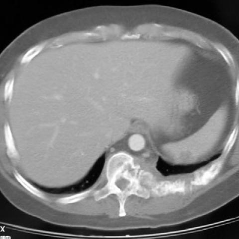
In Septemper 2001, the patient presented with right-sided chest pain. Dual-phase CT of the thorax was performed, and showed a large mass of soft tissue at the back wall of the thoracic cavity, accompanied by osteolytic lesions of the ribs and the vertebrae next to it. There was and extension into the spinal canal. Other, oval-shaped osteolytic lesions with soft tissue masses next to them, were seen on the right side of the chest wall and the left side of the sternum. Lymph-node enlargement was noted at the mediastinum and axillae, especially on the left side. Osteolytic lesions with adjacent soft tissue mass, also occurred on the right side of the sacrum and acetabulum, as CT of that region showed.
In October 2001, core needle biopsy of the mass under CT guidance was performed and pathoanatomic result was "Kaposi's sarcoma". Appropriate therapy was instituted.
In February 2002, CT of the thorax and abdomen was performed to establish the patient's response to the therapy. The osteolytic lesions of the ribs and the vertebrae, seemed to be a little smaller. The lymph nodes of the mediastinum and axillae were smaller as well. The osteolytic lesion at the sacrum was unchanged.
Discussion
Kaposi's sarcoma (KS) is a multifocal neoplasm thought to arise from lymphatic endothelial cells. Histologically, KS is characterised by a proliferation of spindle cells and cleft-like vascular structures. It is the most common neoplasm occurring in HIV-positive patients. It is usually aggressive involving many organs, although skeletal disease is rare. Bone involvement is seen most commonly as invasion of bone from adjacent skin lesions (infiltrative cutaneous or mucosal KS). Primary bone lesions, without involvement of the overlying skin are very rare.
Radiologically, bone KS appears as a periosteal reaction, cortical erosions or as osteolysis. Irregular cortical erosions are most typical, and are probably due to KS arising in the cortex or periosteum. Erosions may also occur secondarily, as a result of external pressure from nodules in the surrounding soft tissue but, in this case, they appeared as smooth lesions. Osteolysis can present as uniform rarefaction, cystic lesions, or almost-complete bone destruction. The possibility of new bone formation with minimal osteoblastic changes has been described in a few instances. These lesions are probably related to the production and release by KS cells of cytokines such as interleukin-1 and interleukin-6, which can stimulate bone resorption, in part by increasing osteoclast development and function. In addition, the pressure generated by enlarging KS nodules may induce resident monocytes to produce cytokines that, in turn, trigger osteoclast activation.
CT scanning more precisely identifies lytic bone changes and MR imaging shows marrow abnormalities similar to those in lymphoma and infection. Soft tissue masses are again more easily identified on MR imaging than CT scanning. Nuclear medicine studies may be useful for further evaluation. Red blood cell pooling in cutaneous lesions can be seen with technetium 99m MDP scans. Combined thallium-201 and gallium-67 imaging reveals a characteristic pattern of uptake with thallium-201, but not with gallium-67. Infection and other neoplasms, such as lymphoma, are typically gallium avid. Nevertheless, biopsy may be necessary for definitive diagnosis.
Differential Diagnosis List
Final Diagnosis
Osseous Kaposi's sarcoma
Liscense
Figures
Osseous Kaposi's sarcoma


Medical Analysis Report
I. Imaging Findings
Based on the provided chest and abdominal CT images and patient information, the following imaging findings are observed:
- Bone destruction: In some areas of the thoracic vertebrae or ribs, there is reduced bone density with focal or patchy osteolytic changes (osteolytic lesion).
- Mild cortical bone erosion or irregular defects are visible in certain regions, suggesting that the lesion may extend outward from the cortical bone.
- Surrounding soft tissue may present masses or nodular changes, although in some affected areas there is no clear indication of skin or mucosal lesions.
- On certain imaging slices, increased soft tissue density or swelling is observed near the lesion, accompanied by possible vascular proliferation.
- No obvious fracture lines are noted, and there are no signs of significant fracture displacement.
II. Possible Diagnoses
Considering the patient’s HIV-positive status, the presence of osteolytic lesions, and the imaging characteristics, the following differential diagnoses should be taken into account:
- Kaposi's Sarcoma: In HIV-positive patients, Kaposi's Sarcoma is among the most common malignancies. Though bone involvement is not frequently seen, it can manifest as cortical bone erosion, osteolytic lesions, or soft tissue masses. Radiologically, it often presents as irregular bone destruction, with or without neighboring soft tissue lesions.
- Bone Lymphoma or Other Malignant Tumors: In the setting of immunocompromise, conditions such as non-Hodgkin’s lymphoma can invade bone and present as bone destruction or osteolytic changes.
- Infectious Bone Disease (e.g., Tuberculosis or Fungal Infections): In HIV patients, opportunistic infections must be considered. Tuberculosis or deep fungal infections may cause chronic osteomyelitis with destructive lesions, requiring further imaging and laboratory tests for differentiation.
III. Final Diagnosis
Taking into account the patient’s age, HIV-positive status, typical osteolytic lesions, and the imaging findings, the most likely diagnosis is Kaposi's Sarcoma involving the bone. Since bone involvement by Kaposi’s Sarcoma is relatively rare, if there is any doubt based on imaging or clinical presentation, pathological puncture or biopsy could be considered for confirmation.
IV. Treatment Plan and Rehabilitation Strategies
1. Treatment Strategy
- Antiretroviral Therapy (ART): Actively controlling HIV viral load to improve immune function is critical for slowing the progression of Kaposi’s Sarcoma.
- Local Treatment: If the lesions are localized and causing significant symptoms, radiation therapy or local injection therapy may be considered upon specialist evaluation to help alleviate bone pain or local destruction.
- Systemic Treatment: For extensively involved lesions affecting multiple bones or other organs, chemotherapy (e.g., taxane-based or anthracycline regimens) or specific therapeutic agents such as α-interferon may be used in combination with ART.
- Symptomatic Management: In cases of bone pain, appropriate analgesics, calcium, vitamin D, and other supportive measures should be provided to reduce symptoms and promote bone health.
2. Rehabilitation / Exercise Prescription Recommendations
Considering the patient’s HIV-positive status and potential bone destruction, rehabilitation programs should be individualized, implemented gradually, and emphasize safety.
- Early Rehabilitation:
- Engage primarily in low-intensity aerobic exercises such as daily walking, cycling, or seated rowing for about 10–15 minutes each session, 3–4 times per week.
- Reduce exercise intensity and duration if pain or fatigue occurs.
- Perform simple range-of-motion exercises and light resistance training, focusing on postural stability and muscle coordination.
- Mid-Stage Rehabilitation:
- If physically capable, gradually increase total aerobic exercise to 20–30 minutes per session, 3–5 times per week.
- Light resistance training can be progressed to moderate intensity (e.g., using small dumbbells or resistance bands), 10–12 repetitions per set, to improve muscle strength and bone support.
- Closely monitor for bone pain or local discomfort to avoid excessive loading.
- Late-Stage Rehabilitation:
- Once the condition is stable and the risk of bone injury is reduced, low-impact exercises such as Tai Chi or yoga can be introduced to further improve balance and muscle endurance.
- In accordance with a physician’s and rehabilitation therapist’s evaluation, increase the intensity of resistance and aerobic training if the bone condition permits.
- Continue with regular follow-ups and medical evaluations, monitoring HIV viral load, hematological indicators, and bone health.
The above exercise recommendations follow the FITT-VP principle: adjusting Frequency, Intensity, Time, Type, Progression, and Volume step by step. For patients with fragile bones or low immunity, safety must be ensured by conducting exercises under the guidance of healthcare professionals.
Disclaimer:
This report is a reference analysis based on limited imaging and medical history information and does not replace in-person consultation or professional medical advice. If you have any further questions or symptoms, please consult a specialist for appropriate guidance.
Human Doctor Final Diagnosis
Osseous Kaposi's sarcoma