Irreducible posterior fracture dislocation of the hip


Clinical History
Moped driver who hit a stationery car sustaining a closed injury to his left hip.
Imaging Findings
The patient, travelling at around 30 miles/ hour on a moped hit a stationery car. He did not have any significant past illnesses. On examination he was haemodynamically stable and his only injury was localised to the left hip. Clinically the left hip was internally rotated and shortened. Sciatic nerve was not affected. Radiographs of the pelvis revealed a posterior fracture dislocation of the left hip (Thompson and Epstein type I) (Fig 1).
Closed reduction under sedation was attempted in the emergency unit. Further radiographs of the left hip revealed that the hip was still dislocated with possible fracture fragment or soft tissue interposition (Fig 2). The patient had a CT of the left hip which showed that the posterior lip fracture of the acetabulum was interposed, preventing the reduction of the hip dislocation (Fig 3). Under general anaesthesia and lateral position through a standard posterior approach the left hip was exposed. Femoral head had button holed through the posterior joint capsule and piriformis. The interposed fracture fragment was reduced without loss of soft tissue continuity following which the hip reduced. The fracture was fixed with two partially threaded cancellous screws (Fig 4). The hip joint was stable in all directions. Post operative recovery was uneventful. Patient was non weight bearing for six weeks and then started on full mobilisation.
Discussion
Hip joint is a well contained ball and socket joint which does not dislocate as easily as the shoulder joint. The preventive mechanism is mainly by the bony contour of the acetabulum which contains the spherical femoral head in association with the labrum, capsule and ligaments. 80 % of hip dislocations are posterior and produced by axial loading of a flexed hip forcing the femoral head out of the acetabulum posteriorly. A pure dislocation occurs when the hip is adducted and a fracture dislocation occurs when the hip is abducted. Associated sciatic nerve palsy (10-14%) can occur as it runs directly posterior to the hip joint. Rarely sciatic nerve palsy can be a late complication when the nerve is encapsulated by heterotopic calcification (1). Quite often it is a neuropraxia that resolves, if not, the prognosis is poor. A significant number of patients, up to 25%, have associated ipsilateral knee injuries (2). Dislocation of the hip has to be reduced within six hours of the injury when the incidence of avascular necrosis is 5% which rises to 50% when the delay is more than six hours. Several traditional techniques have been used to manipulate the posterior dislocation of hip – Bigelow’s, Stimson’s, Allis’ etc., and modifications of these techniques as described by Walden PD (3) and Howard (4). After closed reduction of hip Hougaard et al, recommend computerised tomography to rule out intra articular fracture fragments (5). Avascular necrosis may develop up to 3 years post injury and the incidence is dependent on the time between injury and reduction of the dislocation. Protected weight bearing has no effect on the development of avascular necrosis. When there is no associated posterior fracture dislocation early weight bearing is possible. Heterotopic ossification occurs in 2% after dislocation or fracture-dislocation of the hip, especially when open reduction has been necessary, but it is usually not disabling. Fracture dislocations of the hip are significant injuries with a potential for serious complications both in the immediate future and long term. This case is presented to stress the fact that fracture dislocations of hip may require open reduction within six hours and should be undertaken with care to prevent further soft tissue disruption.
Differential Diagnosis List
Final Diagnosis
Irreducible posterior fracture dislocation of the left hip
Liscense
Figures
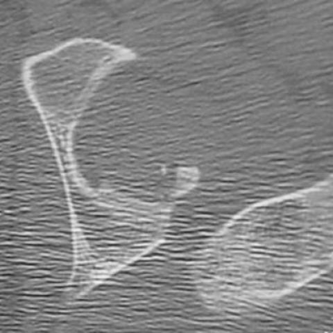
Anteroposterior radiograph of the pelvis

Radiograph following attempted closed reduction

C T scan of left hip
Post operative radiograph of left hip

Medical Imaging Analysis Report
I. Imaging Findings
1. From the provided pelvic AP X-ray, there is a clear abnormal alignment in the left hip region, with the femoral head positioned abnormally relative to the acetabulum, suggesting a posterior direction of dislocation.
2. The acetabular rim shows areas of irregular bone structure or suspicious fracture fragments, indicating a possible fracture of the posterior acetabular rim.
3. Lateral X-ray and CT images demonstrate the femoral head dislocated from the acetabulum and displaced posteriorly and superiorly, with some fracture fragments surrounding the hip joint, further supporting a diagnosis of posterior fracture-dislocation of the hip.
4. Postoperative imaging reveals the presence of internal fixation devices (screws or fixation materials around the acetabulum) used to fix the fracture fragments, and the femoral head is restored to a better position compared to before, with improved alignment.
II. Potential Diagnoses
-
Posterior Hip Dislocation (With or Without Acetabular Fracture)
Cause: In high-speed collisions or high-energy trauma, the hip joint, when subjected to axial loading in flexion and slight adduction, is prone to posterior dislocation. If the force is severe, it may also involve a fracture of the posterior acetabular rim or the femoral head/neck. -
Simple Hip Dislocation
Although the primary presentation is a posterior hip dislocation, if the acetabular fracture fragments are not prominent or do not involve the weight-bearing area, a simple dislocation can be considered. However, X-ray and CT findings in this case indicate involvement of the posterior acetabular rim, thus making a simple dislocation less likely. -
Other Hip Fractures (e.g., Femoral Neck Fracture, Intertrochanteric Fracture, etc.)
These conditions often appear on imaging as a disruption in the proximal femur, usually showing fracture lines in the femoral head-neck region or trochanteric region. Based on the imaging characteristics here, these fractures are clearly distinguishable and not the main consideration.
III. Final Diagnosis
Based on the patient’s young age (18 years), history of trauma (traffic accident), clinical symptoms (hip pain and restricted movement), and imaging findings (posterior dislocation with visible posterior acetabular rim fracture fragments), the most likely diagnosis is:
Posterior Fracture-Dislocation of the Left Hip
In emergency situations, prompt reduction (preferably within 6 hours) is recommended to reduce the risk of avascular necrosis of the femoral head. If there are significant fracture fragments or the reduction is unstable, emergency surgical intervention may be necessary.
IV. Treatment Plan and Rehabilitation
1. Treatment Plan
- Emergency Closed Reduction or Open Reduction: For posterior hip dislocation, the initial approach should be closed reduction under general anesthesia or sedation and analgesia. If closed reduction fails or imaging reveals large fracture fragments impacted in the joint cavity or collapse of the articular surface, open reduction and internal fixation (ORIF) should be considered.
- Intraoperative Assessment of the Joint and Exclusion of Neurovascular Injury: Because the sciatic nerve is located posteriorly, close monitoring of sciatic nerve function, including sensation and motor function, is required during and after surgery.
- Postoperative Imaging Evaluation: Following surgery, X-ray or CT examinations are necessary to confirm the quality of reduction and fixation of fracture fragments, and to exclude any residual fragments or articular incongruity.
- Prophylactic Medications and Other Measures: Where appropriate, consider using low-molecular-weight heparin to prevent deep vein thrombosis, and initiate early distal limb circulation and nerve rehabilitation exercises. In the presence of high-risk factors, prophylaxis against heterotopic ossification (e.g., with NSAIDs) may also be warranted.
2. Rehabilitation and Exercise Prescription
Rehabilitation must follow the principle of gradual progression (FITT-VP) and be tailored to the patient’s individual condition, age, and severity of injury.
-
Early Phase (0–4 weeks post-surgery):
- Absolute immobilization or partial weight-bearing: Depending on the fracture fixation, the physician will typically recommend reducing or avoiding weight-bearing during the initial postoperative period, using crutches or a walker.
- Range of motion exercises: Under minimal or non-weight-bearing conditions, perform passive or assisted active range-of-motion exercises to prevent joint stiffness.
- Isometric muscle contractions: Encourage isometric exercises of the quadriceps, gluteal muscles, etc., to maintain muscle strength.
-
Intermediate Phase (4–8 weeks post-surgery):
- Gradual increase in weight-bearing: After radiographic confirmation of fracture healing progression, weight-bearing can be gradually increased, for example from 20% → 50% → 80%.
- Lower limb strength training: Exercises such as straight leg raises, bridging, and seated ankle pumps can strengthen the muscles around the hip, knee, and ankle.
- Flexibility training: Slowly increase the range of motion of the joint and perform dynamic stretching exercises for the soft tissues around the hip.
-
Late Phase (8 weeks post-surgery and beyond):
- Full weight-bearing and gait training: Once the fracture is essentially stable, transition gradually to full weight-bearing and use walking aids (walker or crutches) to correct gait.
- Return to functional activities: This includes squat training, light jogging, or basic exercises specific to certain sports (like basketball or running); these should be introduced gradually under professional supervision.
Throughout rehabilitation, it is important to have regular follow-up to evaluate hip function, bone healing, and the status of tendons and muscles. If severe pain, swelling, or neurological symptoms occur, contact the attending physician promptly and adjust the plan accordingly.
Disclaimer: This report is based on the provided history and imaging data for reference only and does not replace in-person consultation or professional medical advice. Specific diagnosis and treatment must be guided by clinical evaluation and specialist recommendations.
Human Doctor Final Diagnosis
Irreducible posterior fracture dislocation of the left hip