

Complained mostly of sensory disorders of the leg and foot region.
The patient complained of numbness of the right tibia and foot, and plantar muscle weakness during the past two years. He had no previous history of trauma. Physical examination was significant for sensory loss deficit in the territory of S1 nerve root and reduction of the Achilles tendon reflex, while there were no signs of muscle weakness of the ipsilateral foot. Electromyogram (EMG )was normal.
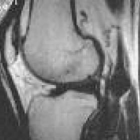
A MRI examination of the knee was performed with a whole body MR scanner (0,5 T), using T1-weighted (TR/TE,300-600/15) images before and after i.v. infusion of contrast medium and gradient echo images (TR/TE 550/22; flip angle 30 degrees), in axial, coronal and sagittal planes.
A round shaped, well-defined mass with smooth margins was identified at the medial aspect of the popliteal fossa, along the tibial segment of the ischial nerve. On T1-weighted images (fig. 1a), the lesion had low signal intensity but on T1-weighted post contrast (fig. 1b), and gradient echo pulse sequences (fig. 2), it had high signal intensity with a central region of low signal, mimicking a target. There were no signs of muscle atrophy or other concomitant findings. The appearance of the mass was identical to neurofibroma which that was proved by histological examination.
The majority of Peripheral Nerve Sheath Tumors (PNSTs) arises from the proliferation of the active and versatile Schwann cells and includes the benign, neurilemmoma (schwannoma) and neurofibroma and the malignant schwannoma. The benign PNSTs constitute 10%-12% of all benign tumors of soft tissues. The most frequent of them is neurofibroma which represents about 5%-6% of all benign tumors of soft tissues with an incidence, in the knee area, up to 8%. It is usually seen during the third decade with no particular predilection between males and females. The mode of neurofibromas formation includes proliferation of endoneural myxomatous matrix, which progressively separates myelinated and non-myelinated axons. Collagen and Schwann cells also proliferate and become embedded in the unorganized intercellular material.
Morphologically, it can be subdivided into three types:
- solitary (the most common type),
- diffuse and
- plexiform.
Neurofibromas can occur as a manifestation of von Recklinghausen disease, especially the plexiform type. It is estimated that only 10% of patients with neurofibromas have Neurofibromatosis I (NF I). Except for those in the skin, neurofibromas may undergo malignant transformation, especially in NF I, and therefore should be excised. Clinical differentiation of benign and malignant PNSTs can be difficult because malignant tumors can quite often be either asymptomatic or cause mild symptoms. A delay in their diagnosis adversely affects prognosis.
Generally, pain at rest, neurologic deficits, short duration of symptoms, large size, prominent vascularity or heterogeneous enhancement and invasive pattern at the margins of the lesion in imaging modalities (U/S, CT and MRI), suggest malignancy.
MRI is the first choice examination for the accurate localization and diagnosis of PNSTs, due to its large field of view and multiplanar capabilities.
The “target sign”, which is mostly seen in T2-weighted images, was histologically found to represent either myxoid tissue encircling cellular matrix containing Schwann cells, fibroblasts, and perineurial cells or myxoid tissue encircling collagen fibers or myxoid tissue encircling a mixed collagen and cellular matrix (fig. 2). Its identification should always raise the suspicion of a neurogenic tumor. It is usually seen in cases of neurofibromas and - much less frequently- in malignant PNSTs. It is useful for differentiating neurofibromas from malignant PNSTs in patients with neurofibromatosis, but its specificity is lower in patients with solitary nerve tumors. Although there are not many types of tumors of peripheral nerves, there is a great variety in their appearance and behavior. Only careful clinical and imaging examinations can lead to differentiation between most benign and malignant peripheral nerve tumors.
Neurofibroma of the ischial nerve


Based on the provided MRI images and the patient’s medical history (42-year-old male with primary complaints of sensory disturbances in the calf and foot regions), the following imaging characteristics are observed:
Taking into consideration the patient’s age, clinical symptoms (sensory disturbance), and MRI signal characteristics—particularly the presence of a “target sign”—the following potential diagnoses can be considered:
These three diagnoses share a common pathological mechanism of nerve sheath cell proliferation; definitive differentiation requires correlation with pathological examination.
In light of the patient’s age (42), the absence of a typical neurofibromatosis history (no indication of NF I), the lesion near the knee joint, the “target sign” on MRI, primarily sensory disturbance, and no obvious evidence of malignancy, the most likely diagnosis at this point is:
If necessary, further confirmation can be sought through the following:
Given that the patient’s primary complaint is sensory disturbance and benign peripheral nerve sheath tumors carry some risk of malignant transformation, the following considerations should be integrated into treatment and rehabilitation:
This report provides only a reference analysis and does not replace in-person consultation or the advice of a medical professional. Patients should consult with a specialist or healthcare team to develop a final individualized diagnostic and rehabilitation plan based on their specific condition.
Neurofibroma of the ischial nerve