Patellar ligament rupture


Clinical History
The patient stumbled and fell on his left knee following which he was unable to extend his left knee.
Imaging Findings
The patient, a company director, stumbled and fell on to his left knee. The patient attended Casualty with complaints of swelling, pain and inability to move his left knee. There was no significant past illness. Patient’s weight was 106 Kg. On examination of the left knee there was diffuse swelling with tenderness and a palpable gap in the patellar ligament. Patient was not able to raise his left leg. Rest of the examination was unremarkable.
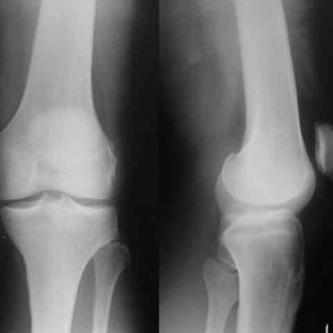
Radiographs of the left knee (Fig 1) revealed a high riding patella with no fractures.
Under general anaesthesia and tourniquet control through a longitudinal midline incision a tension relieving wiring was performed with drill holes through the tibia below the tibial tubercle and through the patella (Fig 2a). While drilling the patella the drill bit broke, this was not explored and left behind (Fig 2b). The patellar ligament was ruptured across its mid substance with the tear extending on both sides for about 3 to 4 cm as shown in the line diagram (Fig 3). The patellar ligament was debrided and repaired with a 5 No. polyester suture and a 00vicryl suture. The suturing and the wiring were such that flexion of the knee up to 90° did not produce any tension in the tendon repair. The extensor retinaculum was repaired with 1 vicryl. The wound was closed in layers. Post operatively the knee was immobilised in a cylinder cast for 6 weeks and radiographs out of plaster showed that the wire had snapped near the tibial entry site (Fig 4). Then patient was allowed supervised mobilisation of the knee up to 90°. At 10 weeks follow up the wound healed well (Fig 5) and the extensor mechanism of the left knee was intact with no extensor lag (Fig 6a) and flexion to 100° was possible (Fig 6b). Patient underwent further physiotherapy and at 6 months follow up had almost full range of movements with good recovery of strength in quadriceps.
Discussion
Patellar ligament injuries are uncommon and occur in the younger patients (1) unlike quadriceps ruptures that occur in the elderly. In the normal patellar ligament, enormous amount of energy is required to rupture it (2). In systemic illnesses like rheumatologic disease, renal failure, and diabetes mellitus patients can present with atraumatic patellar ligament ruptures that is sometimes even bilateral (3). In the absence of a high degree of suspicion and understanding of the condition patellar ligament ruptures can be missed (4).
In patellar ligament ruptures patella is superiorly displaced. This position is called patella alta which is seen on lateral radiograph of the knee. Insall-Salvati ratio (ratio of the greatest length of the patella to the length of the patellar tendon on lateral radiograph) is less than 0.80. In some cases further investigation in the form of ultrasound examination or magnetic resonance imaging may be required.
In acute traumatic patellar ligament ruptures, the treatment is surgical with repair of the ligament which is protected by a ‘tension relieving’ wire fixation through patella and the tibia just distal to the tibial tubercle.
As this case demonstrates early controlled mobilisation of the knee provides satisfactory range of motion and muscle strength.
Complications of surgical management include re rupture, infection, wire breakage, stiffness of the knee.
Differential Diagnosis List
Final Diagnosis
Left Patellar ligament rupture
Liscense
Figures
Radiographs of left knee
Post operative radiographs of left knee


Diagram of the patellar ligament rupture

Radiographs of left knee at 6 weeks post surgery

Figure showing anterior aspect of left knee post surgery

Range of movements at 6 weeks post surgery


1. Radiological Findings
On the anteroposterior (AP) and lateral X-ray views of the patient’s left knee joint, the following main features are observed:
- Patella is positioned higher (i.e., “patella alta”), suggesting possible elongation or rupture of the patellar tendon.
- Post-repair images show “tension band” wire fixation at the lower pole of the patella and the proximal tibial tuberosity, serving to protect the repaired patellar tendon and maintain tension.
- Changes in the local soft tissue density, consistent with postoperative changes and surrounding soft tissue swelling.
- No obvious fracture lines detected, and the joint alignment appears acceptable.
2. Potential Diagnoses
Combining the clinical history (inability to extend the knee after a fall) and radiological findings (patella alta, postoperative fixation), the following diagnoses may be considered:
- Complete or partial patellar tendon rupture: This is a typical cause of inability to actively extend the knee. X-ray findings can indicate a high-riding patella, and metallic tension band fixation may be present post-repair.
- Patellar fracture with associated tendon injury: If the patella was severely traumatized, a fracture could also occur. However, no clear fracture line is visible in this case, and the findings lean more toward a tendon injury.
- Other peri-knee tendon/ligament injuries: For instance, rupture of the quadriceps tendon can likewise lead to extension difficulties, but the marked patella alta here is more indicative of patellar tendon rupture.
3. Final Diagnosis
Considering the patient’s age, history of trauma (fall), clinical presentation (inability to extend the knee), and radiological findings (postoperative tension band fixation, patella alta), the most likely diagnosis is:
Left Knee Patellar Tendon Rupture (Postoperative Repair)
4. Treatment Plan and Rehabilitation Program
Treatment Plan
- Surgical Repair: For acute complete rupture of the patellar tendon, surgical suture and tension band fixation is a common and effective treatment approach.
- Postoperative Immobilization: Early immobilization using a brace or cast is necessary to maintain knee joint stability and prevent re-injury or re-rupture of the tendon.
- Pain and Infection Management: Pain control medication and antibiotics (if infection risk is present) may be provided. Regular follow-up of wound healing is advised to prevent complications.
Rehabilitation Program and Exercise Prescription
Rehabilitation should follow a gradual approach (FITT-VP) and be tailored to the individual patient. The stages can be divided broadly as follows:
-
Early Protection Phase (0-2 weeks after surgery):
- Immobilize the knee in full extension using a brace or splint, with passive knee flexion-extension assisted by healthcare professionals.
- Perform ankle movements and isometric contractions of the quadriceps to help prevent muscle atrophy without increasing joint stress.
-
Progressive Activity Phase (2-6 weeks after surgery):
- Under the guidance of a physician or physical therapist, gradually increase the range of motion in the knee. If allowed, slowly introduce passive and active knee flexion exercises, taking care not to overstretch the repaired tendon.
- Introduce moderate strength training for the quadriceps, hamstrings, and calf muscles, with progressive weight-bearing exercises.
- Frequency of training: 3-5 times per week, with intensity and duration adjusted according to individual tolerance.
-
Strengthening Phase (6-12 weeks after surgery):
- Gradually progress to single-leg weight-bearing and resistance exercises (e.g., light squat or straight leg raises with resistance).
- Adjust the intensity of training appropriately, closely monitoring for pain or swelling.
- Continue regular range of motion exercises and core muscle training.
-
Functional Recovery Phase (3 months post-surgery and onward):
- When appropriate, reintroduce low-impact exercises (e.g., stationary cycling, swimming) to improve knee stability and coordination.
- If functional recovery is good, gradually resume walking, jogging, or low-intensity sports under professional guidance.
Throughout rehabilitation, closely observe knee pain, swelling, and muscle strength. If severe pain, loss of function, or other complications arise, seek medical advice or re-evaluate the rehabilitation plan promptly.
Disclaimer:
This report is a reference medical analysis based on the provided imaging and clinical history. It cannot replace in-person consultation or professional medical diagnosis and treatment advice. The patient should seek further examination and individualized treatment under the guidance of a specialist.
Human Doctor Final Diagnosis
Left Patellar ligament rupture