Segond fracture


Clinical History
Swollen and tender left knee after a fall while skiing.
Imaging Findings
The patient sustained twisting injury to the left knee during a fall while skiing. The knee immediately became swollen and painful. Initial radiograph of the knee showed a subtle avulsion fracture of the left tibia just distal to the lateral tibial plateau and joint effusion. Patient was referred to fracture clinic the following week. On clinical examination, Orthopaedic surgeon suspected an ACL tear and sent the patient for MRI scan of the left knee. Subsequently arthroscopy was performed, which confirmed the full thickness ACL tear.
Discussion
The Segond fracture is a subtle vertical avulsion fracture involving the proximal tibia immediately distal to the lateral plateau. The mechanism of injury is internal rotation and varus stress, which commonly believed to cause abnormal tension on the central portion of the lateral capsular ligament. However, Campos et al have proposed that stresses transmitted via the Iliotibial Tract and the Anterior Oblique Band of the fibular collateral ligament are relevant in the pathogenesis. The importance of a Segond fracture is that it is commonly associated with meniscoligamentous injury, namely:
·Tear of the anterior cruciate ligament (75-100%).
·Menisci Injuries(66-70%).
·Avulsion fracture of the Gerdy tubercle.
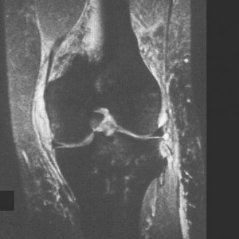
At routine radiography, the avulsed cortical fragment is best seen on the straight anteroposterior view of the knee. By using magnetic resonance ( MRI ) imaging, marrow oedema is seen at the site of the avulsed cortical fragment, but the low signal fragment may be very difficult to appreciate.
Differential Diagnosis List
Final Diagnosis
Segond Fracture
Liscense
Figures
Segond Fracture

Anterior Cruciate ligament tear


Bone edema
Medical Imaging Analysis Report
1. Imaging Findings
This imaging includes routine knee joint X-rays and MRI scans. The X-ray anteroposterior (AP) view shows a small avulsion fragment near the proximal lateral edge of the lateral tibial plateau, appearing as a small, vertically oriented fragment. Additionally, MRI findings in the same region of the proximal lateral tibial cortex reveal localized bone marrow edema and partial soft tissue swelling. MRI suggests possible intra-articular soft tissue injuries, particularly raising concern about the integrity of the anterior cruciate ligament (ACL) and the lateral supporting structures.
2. Potential Diagnoses
-
Segond Fracture (lateral tibial avulsion fracture)
Typically caused by internal rotation and valgus/varus stress on the knee or excessive stress on the lateral tibial attachment, leading to an avulsion at the site where the lateral capsule/ligaments attach. Radiologically, it presents as a small bony fragment avulsed from the proximal lateral tibial plateau. -
Concomitant ACL or Meniscal Injury
Segond fractures frequently occur in conjunction with ACL tears (at a relatively high rate). The meniscus (particularly the lateral meniscus) may also be torn or damaged. Further evaluation with MRI is needed for confirmation. -
Other Tibial Plateau Fractures or Cartilage Damage
Once knee joint biomechanics are altered, other types of plateau fractures or cartilage damage may occur. However, the imaging findings primarily indicate an avulsion fragment, making other forms of tibial plateau fractures relatively less likely.
3. Final Diagnosis
Based on the patient’s history of trauma (a skiing fall with external impact), the imaging findings (a small avulsion fragment from the proximal lateral tibial plateau, localized bone marrow edema), and common associated injuries, the most likely diagnosis is a Segond fracture (with potential ACL or lateral supporting structure injury). A comprehensive MRI evaluation of the ligaments and menisci or an arthroscopic examination is recommended to confirm the extent and nature of soft tissue injuries.
4. Treatment Plan and Rehabilitation Strategy
Treatment Strategy:
· Conservative Treatment: For patients with a small fracture fragment, no significant collapse of the articular surface, and intact or only mildly injured major ligamentous structures (e.g., ACL), bracing, local cold therapy, anti-inflammatory and pain medications, followed by rehabilitation exercises once pain subsides, may be appropriate.
· Surgical Treatment: If there is a severe ACL or meniscal tear, knee joint instability, or if the fracture fragment compromises the articular surface, arthroscopic ligament reconstruction, meniscal repair, and debridement of the avulsed fragment may be considered.
Rehabilitation/Exercise Prescription Recommendations (FITT-VP Principle):
1) Early Stage (Acute Phase, approximately 2-4 weeks post-injury or postoperative):
- Frequency (F): Basic rehabilitation exercises 2-3 times per day.
- Intensity (I): Focus on range-of-motion (ROM) training and isometric quadriceps contractions, staying below the pain threshold.
- Time (T): 5-10 minutes each session, adjusted according to patient tolerance.
- Type (T): Perform active ROM exercises (knee flexion/extension) with a knee brace if needed, along with muscle strengthening, avoiding excessive weight-bearing.
- Progression (P): Gradually increase ROM and strengthening intensity as pain and swelling improve.
- Frequency: 3-4 times per week.
- Intensity: Gradually add weight-bearing exercises, such as single-leg stance balance exercises and low-impact functional training (e.g., light squats, leg presses).
- Time: 20-30 minutes each session.
- Type: May include stationary biking, lower limb coordination, and balance training.
- Progression: Increase power or agility training based on improvements in strength and stability.
- Frequency: 3-5 times per week.
- Intensity: Once knee stability and soft tissue healing are confirmed, progressively increase strength and agility training (e.g., controlled jumping, lateral movements).
- Time: At least 30 minutes each session, adjusted according to the individual's goals and functional recovery.
- Type: Tailored sport-specific training (for a skier, for instance, incorporate ski-simulated drills).
- Progression: Gradually approach normal sporting levels, wearing protective gear as needed and avoiding high-risk movements prematurely.
· Regular follow-ups with a physician or rehabilitation therapist are advised to monitor knee stability and soft tissue healing, and to modify the plan accordingly.
· If pain or swelling increases significantly, reduce or pause training intensity and seek medical advice promptly.
· Maintain balanced nutrition to support bone and soft tissue healing.
5. Disclaimer
This report is a reference analysis based on current imaging and history information. It is not a substitute for an in-person consultation or advice from a qualified physician. The patient should seek further examination and treatment under the guidance of an orthopedic or sports medicine specialist to obtain an individualized treatment plan.
Human Doctor Final Diagnosis
Segond Fracture