Fracture of shaft of little finger metacarpal


Clinical History
The patient sustained an injury to his right hand when he punched a wall.
Imaging Findings
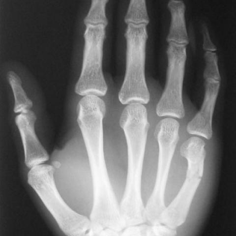
The right hand dominant male, in a fit of anger punched a wall with his right fist. He attended casualty with complaints of pain in his right hand. On examination there was tenderness over the little finger metacarpal. There were no skin wounds, rotational deformity or neurovascular deficits. Range of movements in the little finger joints were reduced due to pain. Radiographs of the right hand revealed a shaft fracture of the little finger metacarpal with a dorsal angulation of 50° (Fig 1).
Under general anaesthesia the fracture was reduced and closed retrograde intramedullary fixation was performed with a 1.8 mm. Kirshner wire. Post operative radiographs showed satisfactory position of the fracture (Fig 2). The right hand was splinted for pain relief, which allowed full mobilisation of the little finger.
Six weeks after operation there was no tenderness at the fracture site. Range of movements was satisfactory with no rotational deformity. Attempts at removal of the Kirshner wire was difficult but with persistence the wire was removed intact (Fig 3). Radiographs after removal of the wire showed satisfactory position of the fracture (Fig 4).
Discussion
Metacarpal shaft fractures are usually caused by direct blow (transverse fractures) and twisting injuries (spiral fractures). Less commonly crushing injuries can cause comminuted metacarpal fractures with significant soft tissue damage. Common problems associated with metacarpal shaft fractures are shortening, angulation and malrotation.
Shortening at fracture site is less common in the middle and ring finger metacarpals due to the deep transverse metacarpal ligaments. Shortening at fracture site produces extensor lag (1).
Most serious of the complications is the malrotation which can significantly affect the hand function while angulation at the fracture site is more of a cosmetic problem. More proximal the fracture more prominent will be the deformity for the same degree of angulation. Dorsal angulation is due to the interosseous muscle action (Fig 5). Angulation at the little and ring finger metacarpals are more acceptable than the one at index and middle finger metacarpals due to more mobile ulnar carpometacarpal joints. Lateral view of the hand is essential to identify the degree of angulation at the fracture site.
Available treatment options include closed reduction and splintage, pinning either transverse (2) or intra medullary (3), plate fixation for multiple metacarpal fractures, external fixation in case of associated soft tissue injury (4). Each has its own advantages and disadvantages (5).
Whatever line of management is chosen the aim should be avoid rotational deformity and stiffness at the metacarpophalangeal and interphalangeal joints.
Complications of treatment of metacarpal shaft fractures include residual angulation, stiffness of joints, non union, pin tract infection, breakage of pins. In the case presented the tip of the pin was bent quite acutely and caused difficulty in its removal. Under these circumstances the possibility of breakage of the tip of the pin and refracture due to overzealous attempts to remove the pin should be kept in mind.
Differential Diagnosis List
Final Diagnosis
Fracture of metacarpal shaft of little finger
Liscense
Figures
Preoperative radiograph of right hand


Per operative radiographs of right hand


Removed Kirshner wire

Radiographs of right hand after removal of wire


Interosseous muscles cause dorsal angulation at fracture site

Medical Analysis Report
I. Imaging Findings
Based on the provided right-hand X-ray, the following are observed:
• A clear cortical interruption and poor alignment in the mid-shaft of the fourth or fifth metacarpal (example), suggesting a fracture line.
• Mild angulation at the fracture site, accompanied by partial swelling of the surrounding soft tissue.
• In later images, percutaneous pin fixation (K-wire) is visible; the distal end of the fixing pin shows a noticeable bend.
• Overall, joint alignment remains acceptable, with no obvious fracture displacement or large bony defect.
• No significant abnormalities are noted in other metacarpals, phalanges, or adjacent joints at the proximal and distal ends.
II. Potential Diagnoses
- Metacarpal shaft fracture (“Boxer’s fracture” or other types of metacarpal shaft/neck fracture)
With a history of punching a hard object, fractures are commonly seen in the fifth or fourth metacarpal shaft/neck. They present as transverse or short oblique fracture lines, often with mild angulation and possible soft tissue swelling. - Other metacarpal injuries (e.g., spiral fractures/comminuted fractures)
Although also associated with forceful impact, imaging findings typically show a spiral or comminuted fracture line, or significant displacement and multiple fragments. Large-scale comminution can be ruled out in this case, making it relatively less likely. - Associated soft tissue injuries around the metacarpophalangeal joints
Since the trauma involved a punch, soft tissue or tendon injury around the joints should be considered. However, from the current imaging, the primary issue appears to be the metacarpal fracture itself, with no prominent soft tissue damage signs visible on radiographs.
III. Final Diagnosis
Considering the patient’s youth, the mechanism of injury (punching a wall), X-ray findings, and clinical fixation treatment, the most likely diagnosis is a fourth or fifth metacarpal shaft fracture. Percutaneous K-wire fixation was performed, but a complication occurred with bending of the wire tip during removal.
IV. Treatment Plan and Rehabilitation
1. Treatment Strategy
- Closed Reduction and Percutaneous Pin Fixation: For metacarpal shaft fractures with mild displacement, a common choice is closed reduction followed by percutaneous K-wire fixation to maintain alignment and reduce recovery time. Attention must be paid to complications such as pin tract infection and difficulties with pin removal.
- Plate and Screw Internal Fixation or External Fixation if Necessary: In cases of multiple fractures or comminuted fractures with severe soft tissue damage, miniature plate-and-screw fixation or external fixation may be considered.
- Postoperative Monitoring: Patients should have regular follow-up X-rays to check fracture healing and fixation stability; careful pin site care is needed to prevent infection or wire breakage.
2. Rehabilitation/Exercise Prescription Recommendations
Follow the FITT-VP principle (Frequency, Intensity, Time, Type, Volume, and Progression), and gradually progress within safe limits. Suggested phases include:
- Early Phase (During Fixation)
• Frequency: Several times daily, perform gentle range-of-motion exercises of adjacent finger joints (e.g., active flexion and extension of the interphalangeal joints).
• Intensity: Minimal, pain-free movements to avoid disruption or loosening of the fixation.
• Time: 2-5 minutes per session, repeated multiple times.
• Type: Mainly passive or assisted joint movements to enhance circulation and prevent joint stiffness.
• Note: Maintain stable fixation and proper pin site care to prevent infection or wire displacement. - Mid Phase (1-2 weeks after pin removal)
• Frequency: 3-4 sessions per week.
• Intensity: Within tolerable pain limits, increase finger flexion/extension and light grip exercises using a soft grip ball or resistance band with mild tension.
• Time: 15-20 minutes per session, gradually increasing.
• Type: Multiple-joint active and coordinated movements to improve muscle flexibility and endurance; progress gradually without rushing into high-intensity weight-bearing exercises.
• Progression: Increase training intensity slowly based on fracture healing status and tolerance to joint mobility. - Late Phase (3-6 months recovery)
• Frequency: 3-5 sessions per week of systematic hand function training.
• Intensity: Gradually returning to near-normal grip strength and finger joint range of motion, allowing some weight-bearing exercises, such as using a grip strengthener or moderate resistance with elastic bands.
• Time: 20-30 minutes per session.
• Type: Incorporate functional training like wringing towels, writing with a pen, and gentle gripping of objects for daily activity simulation.
• Progression: Gradually resume preoperative or normal activity levels; increase load and resistance as patient function improves.
• If the patient has weakened bone quality or other systemic limitations, an individualized program under a professional therapist or physician is advised to prevent re-injury.
Disclaimer: This report is for reference purposes only and cannot replace in-person consultation or professional medical advice. If you have any questions or if your condition changes, please consult a qualified healthcare provider promptly.
Human Doctor Final Diagnosis
Fracture of metacarpal shaft of little finger