Sacral Chordoma


Clinical History
A 71-year-old female patient presented with a four-month history of increasing difficulty with defecation and micturition and a swelling around her right elbow. On examination, it was found that the swelling was fixed to deep tissue.
Imaging Findings
A 71-year-old female patient presented with a four-month history of increasing difficulty with defecation and micturition and a swelling around her right elbow. On examination, it was found that the swelling was fixed to deep tissue. Her past medical history included the diagnosis of a sacral chordoma made eleven years earlier. This had been treated with subtotal excision and radiotherapy. Nine years later, she had represented with further bladder problems, and the imaging study performed at that time had revealed the chordoma to have locally recurred (Fig. 1). A further de-bulking operation had been performed. On this presentation, an MRI and a CT scan of her sacrum and arm were performed (Figs. 2–4) . The results of these findings showed that the sacral chordoma had once again increased in size, with an evidence of more bone destruction. The swelling in the arm was shown to be a large soft-tissue mass involving the triceps muscle with multiple fluid and solid compartments. Radiologically, it had similar features to the sacral chordoma. The differential diagnosis of this mass included a soft-tissue sarcoma or a metastasis from the chordoma. The subsequent histological study confirmed the latter.
Discussion
Chordomas are rare, generally slow-growing malignant tumours, with 50% of them originating in the sacrum. Most sacral chordomas present with pain and at diagnosis are locally extensive which, given the proximity of vital structures, make the treatment options challenging and controversial. These include total excision, subtotal excision, radiotherapy and chemotherapy. Some studies advocate, where possible, radical surgery, which may increase the symptom-free interval before relapse. However, in many tumours this may not be surgically possible, and in addition it carries with it a high risk of morbidity. Therefore, a subtotal debulking procedure is often carried out. The adjuvant radiotherapy procedure remains controversial and can be used pre- or post-operatively, which may increase the remission period. The chordomas can commonly recur locally and more rarely with distant metastases. Local recurrence figures are quoted between 19% and 82% with most studies indicating figures towards the latter end of this spectrum. Distant metastases occur in 14%–30% of the cases, most commonly to the bones, the lung and the subcutaneous tissue. However, deposits in the brain, the pericardium, the ovary and the synovium have been described. Metastases to the skeletal muscle are rare and may be misdiagnosed as primary soft-tissue neoplasms. Their incidence is low in both patients with and without known malignancy, and the skeletal muscle itself is thought to be relevantly resistant to both primary and secondary cancer. In our case, the appearance of the metastatic deposit, despite being non-specific, was radiologically similar to the primary chordoma. This case highlights both that sacral chordomas can develop distant metastases, and that rare combinations of disease do exist. However, it is unlikely that the true incidence is known, as many such cases may well go unreported.
Differential Diagnosis List
Final Diagnosis
Recurrent sacral chordoma, metastatic to the skeletal muscle.
Liscense
Figures
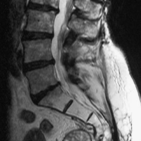
A sagittal T2W MRI scan of the lumbar spine
A sagittal T2W MRI of the lumbar spine and an axial CT scan of the sacrum (bone windows)


An axial T2W MRI at the level of the mid shaft of the humerus

A sagittal T1W post-contrast scan through the humeral mass

Imaging Findings
Based on the provided MRI and CT images, the local bone in the sacral region shows destructive changes, and the lesion exhibits mixed signals with indistinct boundaries, partially involving adjacent soft tissues, suggesting an invasive lesion. Considering the patient's complaints (difficulty with defecation and urination), the sacral lesion may be compressing and invading the surrounding tissues and nerves in the pelvis. In the MRI of the right elbow, a lesion is observed that is connected to deeper structures, with signal characteristics similar to the primary sacral lesion, raising the possibility of metastasis or multifocal disease.
Potential Diagnoses
- Sacral Chordoma (Chordoma) with Distant Metastasis: Sacral chordoma is a rare malignant tumor, often presenting with bone destruction and invasion into surrounding soft tissues. It grows slowly yet may metastasize to distant sites, with MRI revealing similar signal characteristics.
- Other Primary Malignant Sacral Tumors or Metastases: Such as metastatic bone tumors or rare osteosarcomas. However, in light of the findings at the patient’s initial presentation and chronic progression, chordoma is more likely.
- Suspected Soft Tissue Sarcoma (Right Elbow): If the lesion in the right elbow is a solitary soft tissue tumor, primary soft tissue sarcoma should also be considered. Nevertheless, if the imaging features mirror the known sacral lesion, a diagnosis of metastasis is more plausible.
Based on the typical location of the sacral lesion, its slow growth with invasion of surrounding tissues, and similar signal appearance of the right elbow lesion, chordoma metastasis aligns better with the clinical reality than other diagnoses.
Final Diagnosis
Taking into account the patient’s age, chief complaints (dysfunction in defecation and urination), imaging findings of the sacrum and right elbow, as well as the biological behavior of chordoma, the most likely diagnosis is:
“Sacral chordoma with distant soft tissue (right elbow) metastasis.”
To confirm, a biopsy or surgical specimen may be considered for histopathological or immunohistochemical evidence.
Treatment Plan and Rehabilitation
Treatment Strategies
- Surgical Intervention: If feasible, radical surgical resection (including wide excision of the sacral lesion) may be performed to reduce local recurrence. For the right elbow metastasis, resection or debulking surgery can be considered.
- Radiation Therapy: For lesions not amenable to complete surgical resection or where surgery is not suitable, adjuvant or combined radiation therapy (including conventional radiotherapy or proton/heavy-ion therapy) can help extend disease-free survival.
- Chemotherapy and Targeted Therapy: Chordoma generally shows limited sensitivity to conventional chemotherapy. If conditions permit, specific molecular targeted therapies may be considered after multidisciplinary discussion.
- Palliative and Supportive Care: This includes pain management and nutritional support. For elderly patients, comorbidities and overall quality of life should be taken into consideration.
Rehabilitation and Exercise Prescription
After completing major treatments, an individualized and gradual exercise program should be designed under the assessment of physicians or rehabilitation specialists, focusing on:
-
Basic Recovery Training (FITT-VP Principle):
Frequency: 2–3 times per week;
Intensity: Avoid significant pain or fatigue;
Time: 15–30 minutes per session, gradually increasing based on tolerance;
Type: Choose safer exercises in seated or supine positions, including stretching and light resistance training, accompanied by breathing exercises;
Progression/Volume: Increase exercise duration or mild resistance gradually once the patient can complete the current intensity stably. - Muscle Strength and Stability Training: Target the muscle groups around the surgical area or lesion site to mitigate muscle atrophy and decreased joint mobility due to limited activity. Avoid excessive loading and proceed under professional guidance.
- Avoid Repetitive Injury: If there is postoperative pain or sensory disturbance in the sacral region or lower limbs, avoid large forward flexion or twisting motions. For an elbow surgical site, protect the local joint function during recovery.
- Regular Follow-up and Adjustments: As tolerance improves, patients should be reassessed and re-examined to dynamically evaluate tumor control and rehabilitation progress, with exercise plans intensified or adjusted as needed.
If bone fragility or pain is present, extra caution is required. Early in recovery, gentle movements may be performed with support devices or medical personnel assistance, gradually transitioning to independent activity.
Disclaimer: This report is a preliminary analysis based on current information and should not replace in-person consultation or professional medical advice. For any concerns, please seek advice from specialists in orthopedics, oncology, and rehabilitation medicine.
Human Doctor Final Diagnosis
Recurrent sacral chordoma, metastatic to the skeletal muscle.