Bilateral Avulsion Fractures of Plantar Fascia Insertion to Os Calcis


Clinical History
This lady presented at the age of 51 with right hip pain. The hip symptoms seemed to be related to impingement of the labrum.
Imaging Findings
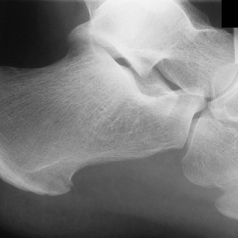
This twenty eight year old man was working on a building site when he fell fom about six feet landing with his feet across an upturned plank of wood. The plank was approximately at the level of his metatarsal heads but his heels were able to keep moving downwards forcibly dorsiflexing both feet. He then found weightbearing difficult and was taken to the Emergency Department. Plain radiographs were taken of both os calcis which showed avulsion fractures of the plantar fascia insertion.
Discussion
Avulsion fractures of the plantar fascia insertion are uncommon. In this case the patient was managed with simple analgesia and mobilised with crutches for two weeks until his pain settled down. Review at a later date showed him to have no long term disability from his injury.
Differential Diagnosis List
Final Diagnosis
Bilateral Avulsion Fractures of Plantar Fascia Insertion to Os Calcis.
Liscense
Figures
Right Os Calcis Plantar Fascia Insertion Avulsion Fracture
Left Os Calcis Plantar Fascia Insertion Avulsion Fracture

Radiological Analysis Report
I. Radiological Findings
Based on the provided foot X-ray (lateral view), the calcaneus (heel bone) appears generally intact. However, in the region of the plantar fascia insertion (at or near the medial tubercle of the calcaneus), there is a suspicious slight avulsion-like change. The cortical margin in this area appears irregular, although there is no significant soft tissue swelling. Combined with the textual description, this suggests a possible avulsion fracture at the plantar fascia insertion. Overall bone quality appears relatively uniform, with no obvious signs of osteoporosis or bone defects. The alignment of the ankle joint is essentially normal, and there is no notable abnormality in other articular surfaces. Further magnification or careful examination may help confirm the size and location of any avulsed fragment, aiding in assessing the extent of avulsion and soft tissue involvement.
II. Potential Diagnoses
- Avulsion Fracture of the Plantar Fascia Insertion
This type of fracture, while not very common, can be related to sports injuries or trauma. On X-rays, it can manifest as a small separated fragment of bone at the medial side of the calcaneus or near the plantar fascia attachment site, where the cortical bone at the insertion may appear irregular. Patients often experience pain and localized tenderness due to an acute excessive load or pull on the plantar fascia.
- Calcaneal Spur (Changes Associated with Plantar Fasciitis)
In chronic plantar fasciitis, bony outgrowth (commonly referred to as a “heel spur”) can develop at the plantar aspect of the calcaneus due to repeated traction stress. Such changes are more common in middle-aged or older adults, or in cases of recurrent stress injuries. On X-rays, these typically appear as bony protrusions on the bottom of the calcaneus rather than a distinct avulsed fragment.
- Other Types of Calcaneal Fractures (e.g., Compression or Stress Fractures)
High-energy trauma or prolonged repetitive stress can also lead to other forms of calcaneal fractures. However, classic calcaneal fractures often involve collapse of the articular surface or a change in the overall shape of the calcaneus, which differs from the localized avulsion at the plantar fascia insertion seen in this case.
III. Final Diagnosis
Considering the patient’s age (28, male), symptoms (plantar or heel pain), and radiological findings (irregular cortex at the plantar fascia insertion of the calcaneus, with a possible small avulsed fragment), as well as the clinical treatment process described, the most likely diagnosis is an avulsion fracture at the plantar fascia insertion. If any diagnostic uncertainty remains, a CT or MRI could be considered for clearer visualization of the fracture fragment size and the extent of soft tissue damage.
IV. Treatment Plan and Rehabilitation
- Treatment Strategies
- Conservative Treatment: For minimal avulsion fractures with no significant fragment displacement, the main approach is rest, immobilization, reduced weight-bearing, and pain management (e.g., NSAIDs). Depending on the severity of pain, a brace or protective device may be used to support the foot.
- Surgical Indication: If the avulsed fragment is large and significantly displaced, or if there is severe tearing of the plantar fascia leading to functional impairment, persistent pain, or instability, surgery may be considered. This is typically pursued once imaging confirms significant displacement or soft tissue damage.
- Rehabilitation / Exercise Prescription
- Early Stage (0–2 weeks):
- Reduce Foot Load: Use crutches or a walker to minimize weight-bearing.
- Anti-inflammatory and Pain Relief: NSAIDs or topical medications if needed.
- Cold Therapy: 3–4 times a day, 15–20 minutes each time, to control swelling and pain.
- Mid Stage (2–6 weeks):
- Partial Weight-Bearing: As pain subsides, gradually transition to assisted partial weight-bearing with crutches.
- Gentle Plantar Stretching: Perform light stretches of the plantar fascia and posterior calf muscles, within a pain-free range.
- Range of Motion Exercises: Increase active and passive ankle range of motion, such as dorsiflexion/plantarflexion and inversion/eversion.
- Late Stage (6 weeks and beyond):
- Gradually Increase Weight-Bearing and Walking: Once pain is largely resolved, progressively discontinue crutches and braces to restore normal weight-bearing.
- Strength Training: Use resistance bands or bodyweight exercises to strengthen the calf and foot muscles, progressing gradually to avoid excessive strain or pain.
- Functional Training: Incorporate standing balance, single-leg support, and light jumping (if pain-free) to restore tolerance to load and impact.
- Applying the FITT-VP Principle:
- Frequency: 3–5 sessions per week.
- Intensity: In early stages, avoid aggravating pain at the avulsion site; later phases can see gradual increases in weight-bearing and activity levels.
- Time: 20–30 minutes per session, including stretching, strengthening, and gait training.
- Type: A combination of range of motion exercises, peripheral strength training, and balance/stability work.
- Progression: Advance from non-weight-bearing to partial weight-bearing, then to full weight-bearing, and eventually incorporate agility, speed, and coordination exercises as tolerated.
- Volume: Increase total training volume gradually as rehabilitation progresses.
- Early Stage (0–2 weeks):
V. Disclaimer
This report is based on the provided imaging and clinical history for reference only. It does not replace an in-person consultation with a professional physician or further diagnostic workup. Specific treatment and rehabilitation plans should be determined by a professional medical team, taking into account the patient’s actual condition.
Human Doctor Final Diagnosis
Bilateral Avulsion Fractures of Plantar Fascia Insertion to Os Calcis.