Superficial peroneal nerve schwannoma


Clinical History
Palpable soft-tissue mass of the right leg
Imaging Findings
The patient consults for a swelling upon the lateral malleolus of the right leg, with neuralgic pain. There was no sensitive or motive deficit. Radiographics were normal. Ultrasonography and MRI of the leg were realized.
Discussion
Schwannoma is a rare benign neurogenic tumor developing from schwan cells but it is the most common of all peripheral tumours (1, 2). The risk of malignant degeneration in a schwannoma is exceedingly rare. It occurs at all ages but most commonly in persons between the ages of 20 and 50 years and represents approximately 5% of benign soft-tissue neoplasm (3). Men and women are affected equally. Schwannoma of the foot is very rare (3). Most lesions are solitary and present as a slowly growing painless soft-tissue mass (3). On clinical exam, this mass is often mobile in transverse direction (2), but fixed along the nerve; painful paresthesia in the dermatome of the nerve of origin, similar to tinel’s sign may be present (1, 2). Sonography identified the 3-dimensionnal localization of the nerve tumor its origin from a nerve fascicle, and its relationship with non involved nerve fascicles (4). MRI is the best imaging investigation for the diagnosis of schwannoma. On T1-weighted images, schwannomas have intermediate signal intensity similar to that of muscles. Sometimes they are hardly visible. On T2-weighted images schwannomas show markedly increased signal intensity. Cystic lesions within them, which are typically found in schwannomas, show low signal intensity on T1-weighted images and high on T2-weighted. Usually, small schwannomas tend to enhance uniformly after gadolinium whereas larger lesions show a more heterogeneous enhancement. The detection of a capsule on MRI visualized as low intensity rim could be detected in 70% of the schwannomas and in 30% of the neurofibromas (3). The principle of schwannoma surgery is simple enucleation of tumour without damaging the nerve (2, 3). Nerve fibres do not traverse the substance of the schwannoma and operative excision should therefore not compromise nerve function.
Differential Diagnosis List
Final Diagnosis
Schwannoma of the superficial peroneal nerve
Liscense
Figures
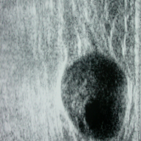
Ultrasonography shows an elongated ovoid mass of 2 cm, with double structure, tissular and cystic. The tumor is eccentric to the nerve trunk of origin
Coronal T1 image of the distal right leg extremity. Schwannoma appears as a fusiform shape oriented longitudinally in the nerve distribution, eccentric to the nerve trunk of origin. It shows low signal intensity.

Sagittal T1 image of the distal right leg extremity. Schwannoma appears as a fusiform shape oriented longitudinally in the nerve distribution, eccentric to the nerve trunk of origin. It shows low signal intensity.

coronal T2 image of the distal right leg extremity. Schwannoma shows heterogeneous high signal intensity.

Post-gadolinium injection coronal T1 image of the distal right leg extremity. Schwannoma shows inhomogeneous enhancement after contrast due to a central cystic degeneration.

Medical Analysis Report
I. Imaging Findings
Based on the provided MRI images, a round or oval lesion is observed within the soft tissue of the right lower leg (proximal to the ankle joint / foot and ankle region), showing relatively well-defined margins. On T1-weighted images, the lesion generally exhibits signal intensity that is moderate or slightly lower than that of muscle, with some areas of slightly low signal. On T2-weighted images, the lesion appears as high signal with clear boundaries; some cyst-like low or high signal zones may be present internally. It is clearly demarcated from surrounding tissues, and a capsule-like low signal ring may be seen on certain sequences. Located near the course of a nerve with a round or oval shape, its overall appearance is consistent with a typical schwannoma (Schwannoma).
II. Potential Diagnoses
- Schwannoma: Based on the MRI findings, a palpable soft tissue lump, patient age, and lesion location, schwannoma is the primary consideration. Its features include slow growth, frequently appearing as an elliptical or round mass, moderate or slightly low signal on T1, high signal on T2, and possible homogeneous or partial enhancement after contrast administration.
- Neurofibroma: Another nerve-origin tumor. Compared to schwannomas, there may be differences in the presence of a capsule and the proportion of cystic changes. Histological or tissue examinations are often required for definitive differentiation.
- Tenosynovial Giant Cell Tumor or Ganglion Cyst: These commonly arise near tendons or tendon sheaths. However, their MRI characteristics often show relatively low or mixed signal on T2, and they do not typically exhibit a clear relationship with nerve structures as neurogenic tumors do.
- Lipoma or Hemangioma: These should also be considered in the differential diagnosis of soft tissue tumors. Nevertheless, lipomas usually exhibit characteristic fatty signals on both T1 and T2, while hemangiomas often show flow void signals. These findings do not match well with the MRI features in this case.
III. Final Diagnosis
Considering the imaging features, the patient's gender and age (49-year-old female), the slow-growing, palpable nature of the mass, and the typical MRI presentation with clear margins and correlation to the nerve pathway, the most likely diagnosis is Schwannoma.
In cases with atypical signs or high suspicion of malignancy, further confirmation via histopathological biopsy or intraoperative frozen section is recommended.
IV. Treatment Plan and Rehabilitation Program
1. Treatment Strategies
- Surgical Treatment: For schwannomas that cause significant symptoms or show progressive enlargement, surgical excision (enucleation) is commonly indicated. Since schwannomas typically arise from the external sheath of the nerve, the nerve fibers are usually pushed aside, allowing complete removal while preserving nerve function.
- Conservative Treatment: If the lesion is very small with mild symptoms, active surveillance under close follow-up may be considered. However, surgery should be considered promptly if notable pain, sensory disturbances, or an increase in mass size are observed.
2. Rehabilitation/Exercise Prescription Suggestions
The main goals of postoperative rehabilitation include:
- Alleviating local swelling and pain, promoting tissue healing.
- Preserving and gradually recovering limb functionality to prevent muscle strength loss.
Planning rehabilitation under the FITT-VP principle (Frequency, Intensity, Time, Type, and Progression):
- Early Phase (1-2 weeks postoperative):
- Frequency: Light activities or gentle ankle joint movements daily or every other day.
- Intensity: Within tolerable pain limits, avoiding excessive weight-bearing or intense exercises.
- Time: 5-10 minutes per session, multiple times a day.
- Type: Elevating the affected limb, ankle pump exercises, and passive or active joint movements to reduce swelling.
- Progression: Increase range of motion gradually according to wound healing progress.
- Mid Phase (2-6 weeks postoperative):
- Frequency: 3-4 times per week.
- Intensity: Mild to moderate; gradually increase joint mobility and muscle strength without exacerbating pain or swelling.
- Time: 15-20 minutes per session.
- Type: Active ankle movements in all directions, light resistance exercises (e.g., using resistance bands), and stability exercises for the lower extremities (e.g., single-leg standing).
- Progression: As pain and swelling decrease, gradually restore a normal gait.
- Late Phase (6 weeks postoperative and beyond):
- Frequency: 3-5 times per week.
- Intensity: Moderate levels, increasing progressively based on individual tolerance.
- Time: 20-30 minutes per session.
- Type: Gradually incorporate functional exercises, such as low-impact aerobic activities (indoor cycling, swimming), comprehensive lower-limb strengthening (e.g., squats, lunges), combined with balance and proprioception training.
- Progression: Emphasize full restoration of normal gait and everyday functional ability. If any nerve symptoms persist, avoid excessive stretching or high-impact exercises.
Throughout rehabilitation, closely monitor surgical site pain, swelling, and neurological function. Any significant deterioration or anomalies require prompt reevaluation and adjustment of the rehabilitation plan.
Disclaimer
This report provides a reference-based analysis rooted in the supplied information and does not replace in-person medical evaluation or professional healthcare advice. The definitive diagnosis and treatment planning should be determined by qualified medical professionals, taking into account the patient’s full medical history, physical examination findings, and other auxiliary test results.
Human Doctor Final Diagnosis
Schwannoma of the superficial peroneal nerve