A case of localised gigantism


Clinical History
A 15 year old boy presented with localised gigantism and pigmentation of the distal right lower limb. Imaging revealed gross localised soft tissue and bony hypertrophy with grossly increased abnormal vascularities and arterio-venous channels.
Imaging Findings
A 15-year-old boy presented with a hugely swollen right leg, a steadily worsening limp and history of a discolored patch on the skin of the affected limb since birth which gradually became darkish in color. There was no other associated illness or injury. Distal right lower limb was hypertrophied below knee extending beyond the ankle with darkish discoloration of the thickened and rough skin over it (Fig 1). Tortuous subcutaneous vascular channels were seen without ulceration or tenderness but local temperature elevation, a fine thrill and a vague murmur could be appreciated on auscultation. A voluminous radial pulse with mild tachycardia was the systemic feature. Plain radiographs of the affected limb (Fig 2) showed bony hypertrophy and bowing with an abnormal alignment of the ankle joint. Chest radiograph and ultrasonography of the whole abdomen revealed normal findings. Doppler evaluation of the affected limb (Fig 3) showed low resistance flow in the femoral, popliteal, anterior and posterior tibial arteries with grossly increased venous flows. No exact arterio-venous connection could be delineated. CT scanogram revealed limb lengthening. Axial plain images showed hypertrophied tibia and fibula with hypertrophy of the muscles of the leg, gross thickening of the subcutaneous tissues and overall increased bulk of the limb extending from below the knee down to below the ankle. Contrast study revealed grossly increased vascularities in the affected region with early venous filling (Fig 4). CT angiogram confirmed the presence of increased vascularities, dilated tortuous vessels and multiple arterio-venous channels (Fig 5-6).
Discussion
The association of arterio-venous fistulae with localized soft tissue and bone hypertrophy, cutaneous capillary malformations of an extremity which could be in the form of a port-wine stain, and congenital venous abnormalities or varicosities is termed as Klippel Trenaunay Weber Syndrome, also referred to by some as the Parker Weber Syndrome,. This is a rare sporadic condition with no racial or geographic predisposition. Although the cause is unknown, many investigators believe the Klippel-Trenaunay-Weber syndrome results from a disturbance in embryogenesis, probably in the third to sixth week of gestation. The bone and soft tissue abnormalities (localized gigantism) occur in the same region as the vascular malformations. Usually only one lower limb is involved in the disease. Historically speaking, in 1900 noted French physicians Klippel and Trenaunay first described a syndrome in 2 patients presenting with a port-wine stain and varicosities of an extremity associated with hypertrophy of the affected limb's bony and soft tissue. In 1907, Parkes Weber, an English physician, unaware of Klippel and Trenaunay's report, described a patient with the three aforementioned symptoms as well as an arterio-venous malformation of the affected extremity. This syndrome affects females and males equally, shows no racial predilection and presents at birth or during early infancy or childhood. Other features that may be seen include lymphatic obstruction, spina bifida, hypospadias, polydactyly, syndactyly, oligodactyly, hyperhidrosis, hypertrichosis, paresthesia, decalcification of involved bones, chronic venous insufficiency, stasis dermatitis, poor wound healing, ulceration, thrombosis, and emboli. Arterio-venous fistulae worsen the prognosis of the disease. Cardiac hypertrophy or high-output congestive heart failure may occur later on. In our case, the patient had a definite history of a stain-like lesion on the affected limb since birth which had gradually turned darkish till it attained the present color. There was definite soft tissue and bony hypertrophy along with the abnormal arterio-venous channels, as is evident from the investigations done. Though other complications as detailed above were not present, the patient did have a hyperdynamic circulation clinically. On these bases the case was diagnosed to be that of Klippel Trenaunay Weber Syndrome. The patient has since been referred to a higher center for assessment to undergo intravascular embolisation of the abnormal vascular channels and reconstructive surgery of the limb.
Differential Diagnosis List
Final Diagnosis
Klippel Trenaunay Weber Syndrome
Liscense
Figures
Fig 1: Localized gigantism involving distal right lower limb

Fig 2: Lateral radiograph showing bony hypertrophy and bowing of both tibia and fibula with an abnormal alignment of the ankle joint.

Fig 4: Axial contrast CT section showing hypertrophied bones and muscles, thickened subcutaneous tissues, overall increased bulk along with grossly increased vascularities and early venous filling.
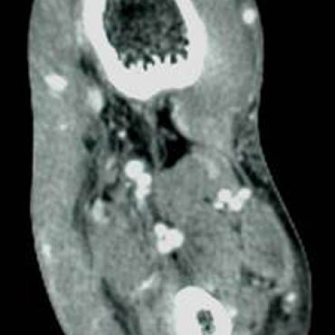
Fig 5: CT angiogram revealing presence of increased vascularities, dilated tortuous vessels and multiple arterio-venous channels.

Fig 3: Comparative depiction of Doppler signals in both common femoral and popliteal arteries along with the spectral waveforms.

Fig 6: SSD reformat of affected limb depicting hypertrophied bones and tortuous vascularities.

Medical Imaging Analysis Report
I. Imaging Findings
Based on the provided images (including X-ray, CT, and vascular ultrasound of the lower limb), as well as the patient’s clinical presentation, the following can be observed:
- The distal lower limb shows significant enlargement, manifesting as thickened soft tissue and bony hypertrophy (“localized giant deformity”), localized to the distal right lower limb.
- The skin surface displays color changes and pigmentation similar to “port-wine stains,” suggesting the presence of capillary and/or venous malformations.
- X-ray shows thickened bone in the affected area, possibly with local deformities, but no definite pathological fractures.
- CT imaging indicates soft tissue overgrowth, abnormal vascular pathways, and low-density lesions in some areas (suggesting vascular dilation or increased vascular channels).
- Vascular ultrasound and angiographic studies (such as MRA/CTA or DSA) demonstrate numerous abnormal arteriovenous channels and venous dilation, with marked hemodynamic alterations.
II. Potential Diagnoses
Taking into account a 15-year-old patient with localized hypertrophy of soft tissues and bones in the right lower limb, accompanying abnormal vascular channels and pigmentation, the following possibilities are considered:
- Klippel-Trenaunay-Weber Syndrome (KTS/KTWS):
Typical features include (1) localized limb hypertrophy (bone and soft tissue), (2) venous or capillary malformations (e.g., “port-wine stains”), and (3) abnormal vasculature such as arteriovenous connections or dilatation. In this case, vascular imaging indicates abnormal arteriovenous channels, along with limb enlargement and cutaneous pigmentation, which is highly representative. - Proteus Syndrome:
This can also present with asymmetric overgrowth of limbs or other body parts, but often involves multiple areas including the cranium and visceral organs, not confined to a single limb. Given that this patient’s main changes are limited to the lower limb and lack the multi-system manifestations typical of Proteus syndrome, it is not currently the primary consideration. - Parkes Weber Syndrome:
Its clinical features are very similar to Klippel-Trenaunay-Weber syndrome, and it can also present with arteriovenous malformations and conical limb enlargement. Some experts view it as part of the same spectrum or a subtype. - Hemangioma or Lymphangioma-Related Syndromes:
Other rare vascular tumor diseases (such as extensive cavernous hemangiomas) may lead to localized limb enlargement, but their distribution typically differs on X-ray and vascular imaging, and the type of skin lesion is usually different.
III. Final Diagnosis
Based on the patient’s age, symptoms (skin spots on the limb present from birth or early childhood, gradually darkening), imaging findings (soft tissue overgrowth, bony hypertrophy, widespread abnormal arteriovenous channels), and clinical hemodynamic changes, the most likely diagnosis is:
Klippel-Trenaunay-Weber Syndrome
To further clarify the vascular pathways and evaluate the risks and benefits of treatment, additional angiographic studies and multidisciplinary collaboration (interventional radiology, vascular surgery, orthopedic surgery) may be considered.
IV. Treatment Plan and Rehabilitation
- Treatment Strategies:
- Interventional Therapy: Employ embolization or sclerotherapy on abnormal arteriovenous fistulas or vascular malformations to reduce blood flow or block abnormal channels, mitigating swelling and improving hemodynamics.
- Surgical Intervention: May involve partial resection/reconstruction of soft tissue or bone to alleviate local deformities and functional impairment. For significant skin and subcutaneous hemangioma-like lesions, appropriate plastic or reparative procedures may be performed.
- Medication: If indicated, consider anticoagulants and compressive therapy (compression stockings, bandages) to relieve edema and prevent thrombosis.
- Observation and Follow-up: If the condition is stable or symptoms are mild, conduct regular follow-ups to monitor limb growth, progression of vascular abnormalities, and cardiovascular function changes.
- Rehabilitation and Exercise Prescription:
The main goals are to promote lower limb function, improve blood circulation, and prevent joint stiffness and potential muscle strength decline. Carefully consider cardiovascular load and the characteristics of the local vascular malformation, proceeding gradually.
- Early Stage (Functional Maintenance):
- Focus on low-intensity exercises such as joint active range of motion while seated or supine, 2–3 times per day, 10–15 minutes each session.
- Depending on tolerance, choose light resistance exercises (using resistance bands or small dumbbells), primarily targeting ankle and knee joint mobility while avoiding increased vascular pressure.
- Intermediate Stage (Muscle Strength Enhancement):
- Gradually increase lower limb resistance training, such as seated knee flexion and extension or light weight-bearing exercises, 3–4 times per week, 20–30 minutes each session.
- Add stationary cycling or water-based activities to aerobic exercise (the buoyancy of water can reduce limb load and improve blood circulation).
- Late Stage (Cardiorespiratory and Functional Training):
- With professional guidance, try moderate-intensity aerobic exercises (e.g., brisk walking, elliptical machines) 3–5 times a week for about 30 minutes each session.
- Emphasize symmetrical strength and flexibility training of the lower limbs, avoiding high-impact activities (like running and jumping) that may stress abnormal vasculature.
- Precautions:
- Closely monitor skin color, swelling, and pain in the lower limb. If pain worsens or swelling becomes more pronounced, reduce activity or pause exercise.
- Carefully assess cardiovascular load to prevent excessive cardiac compensation; conduct cardiac function tests if necessary.
- When wearing compression stockings or bandages, ensure the correct pressure gradient to prevent complications such as thrombosis or ulcers.
- Early Stage (Functional Maintenance):
Disclaimer: This report is for reference only and cannot replace an in-person consultation or a professional physician’s actual diagnosis and treatment recommendations. If the patient experiences any discomfort or questions, please seek medical attention promptly to obtain an individualized diagnosis and treatment plan.
Human Doctor Final Diagnosis
Klippel Trenaunay Weber Syndrome