RADIATION-INDUCED OSTEOCHONDROMATA


Clinical History
Multiple firm lumps gradually increasing in size.
Imaging Findings
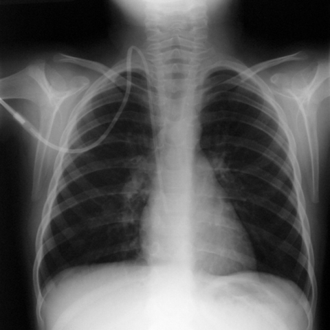
A 16 y.o. boy presented with multiple firm lumps in right middle finger, right ankle and left shoulder with mechanical symptoms. He received total body irradiation prior to bone marrow transplantation 10 years ago aged 5 (1995) for acute lymphoblastic leukaemia which is currently in complete remission. There is no history of other skeletal problems or family history of skeletal disorders. Physical examination demonstrated multiple bony lumps. Plain radiographs were performed of the ankle, shoulder, and right hand.
Discussion
Radiation-induced osteochondromata (RIO) have been reported to be the most common skeletal abnormality after total body irradiation (TBI) in the growing skeleton. The incidence of osteochondromata in the general population is reported at ~1% increasing to ~12% with local high dose radiation therapy. [1].The incidence of RIO varies between 6-24% in previous studies and is directly related to the age at irradiation [2, 3]. In one previous study children who received TBI before 5 years of age developed osteochondromata in 24% of cases compared to no osteochondromata in children irradiated after 5 years of age [2]. In another study, RIO were only seen in children irradiated before 8 years of age [1]. In the study by Fletcher et al, RIO were most common in the long bones and phalanges of the upper and lower limbs. Out of 19 osteochondromata in 5 patients in the upper and lower limbs in this series, the majority (>80%) were at the growing ends of bones, i.e. the proximal humerus, distal radius/ulna, around the knee and epiphyseal ends of phalanges. The distribution in our patient also follows a similar pattern. Although the pathogenesis of RIO is poorly understood, it is probably due to a failure of differentiation of reserve cell layer in the epiphysis and consequent persistence of this undifferentiated cartilage in the metaphysis which later develops into an osteochondroma. Furthermore, chemotherapy and radiotherapy can cause growth retardation resulting in the epiphysis staying open for longer and allowing the osteochondroma more time to develop [2,5]. It was postulated that the development of osteochondromata after TBI may be due to mutation of EXT 1, 2, 3 genes as in hereditary multiple exostoses [1]. Growth hormone treatment in these patients does not seem have an important role in the development of these lesions as shown by Taitz et al [2]. It is difficult to establish the exact latent period after which osteochondromata develop after TBI, because most children are asymptomatic and the lesions are identified casually. However, the time period between the clinical identification of these lesions and the previous irradiation is approximately about 6 years. The minimum radiation dose for the induction of osteochondromata is 1 Gy; most osteochondromata have been reported in patients who received local dose between 15-55 Gy while in another study [3] the dose was 12 Gy. In the general population malignant transformation of solitary osteochondroma into a low grade osteosarcoma is rare, being less than 1%. In multiple exostoses the incidence of malignant degeneration is higher at about 2.5%. There are only three reported cases of RIO undergoing malignant transformation. Thus the exact percentage of transformation into chondrosarcomas or osteosarcomas cannot be deduced, but the degree of sarcomatous change is probably directly related to the dose of radiation.
Differential Diagnosis List
Final Diagnosis
Radiation-induced osteochondromata
Liscense
Figures
Fig. 1.

Fig. 2 (A)

Fig. 2 (B)

Fig. 2 (C)

Fig. 2 (D)

Medical Imaging Analysis Report
I. Imaging Findings
X-ray images from multiple regions (upper limbs, lower limbs, and hands, etc.) show multiple exophytic lesions near the metaphysis or metaphyseal-diaphyseal junction, presenting as distinct bony prominences, with some lesions exhibiting a cartilage cap. The lesions have clear margins and demonstrate a continuity of bony structure, suggesting they are connected to the main bone shaft. No obvious swelling or abnormal soft tissue shadows are seen in adjacent soft tissues. Overall, the location and morphology of these protruding lesions are consistent with the common forms of osteochondroma (exostosis).
II. Possible Diagnoses
-
Radiation-Induced Osteochondromata (RIO)
Given that the patient may have a history of systemic or localized radiation therapy in the past and is currently in a growth phase, there is an increased risk of RIO. The multifocal lesions located near the metaphysis, presenting with an osteocartilaginous protrusion on imaging, align with the distribution and morphologic characteristics of radiation-induced osteochondromas reported in the literature. -
Hereditary Multiple Exostoses (HME)
This condition also presents with multiple osteochondromas, often detected during adolescence. However, HME typically involves a family history or a known genetic mutation, which necessitates further investigation of genetic or familial factors for exclusion. -
Other Benign Osseous or Cartilaginous Tumors
For instance, enchondromas or other rare benign bone tumors can also present with localized bony proliferation or expansion. Nevertheless, the typical pattern of multiple lesions aligns more closely with osteochondromas, making the likelihood of other possibilities relatively lower.
III. Final Diagnosis
Considering the patient’s potential radiation therapy history, age, the characteristic multiple exostotic morphology on X-rays, and the involvement of metaphyses and metaphyseal-diaphyseal junctions, the most likely diagnosis is Radiation-Induced Osteochondromata (RIO). If needed, further imaging follow-ups or MRI evaluations of the cartilage cap thickness may be carried out, and biopsy is recommended at an appropriate time if the lesions exhibit rapid growth or suspicious malignant features, to rule out malignant transformation.
IV. Treatment Plan and Rehabilitation
1. Treatment Strategy
- Regular Imaging Follow-up: For stable lesions without obvious symptoms, periodic (e.g., every 6-12 months) X-ray or MRI exams can be conducted to monitor changes in lesion size, cartilage cap thickness, and surrounding tissue, ensuring early detection of potential malignant transformation or rapid growth.
- Indications for Surgery: In cases where lesions cause pain, neurovascular compression, restricted movement, or are suspected of malignant transformation, surgical excision can be considered. Complete resection of the osteochondroma is typically performed, with bone reshaping as needed to maintain skeletal structure and function.
- Comprehensive Management: If skeletal dysplasia or limb length discrepancies remain, further orthopedic evaluations or procedures may be necessary to correct limb length and improve overall function.
2. Rehabilitation/Exercise Prescription
In the absence of significant pain or deformity, moderate and progressive functional exercises can be carried out. The FITT-VP principle (Frequency, Intensity, Time, Type, Volume, Progression) should be followed:
-
Early Recovery Phase (post-surgery or under observation)
- Frequency: 3-4 times per week.
- Intensity: Low load and low impact exercises, such as walking and simple range-of-motion activities.
- Time: Start at 15-20 minutes per day, gradually increasing as tolerated.
- Type: Focus on flexibility and range-of-motion exercises (e.g., stretching, light functional activities).
-
Mid-Stage Training (stable rehabilitation phase)
- Frequency: 3-5 times per week.
- Intensity: Slightly higher than early phase, adding moderate resistance training (e.g., resistance band exercises, low-weight strength training).
- Time: Approximately 30 minutes each session, possibly divided into segments.
- Type: Maintain joint flexibility while strengthening and stabilizing muscle groups, including the core and main upper and lower limb muscle groups.
-
Late-Stage Training (functional strengthening phase)
- Frequency: 3-5 times per week, including cross-training (e.g., swimming, cycling) to reduce impact.
- Intensity: Gradually increase to moderate or moderately high intensity as appropriate for the patient, avoiding excessive load.
- Time: 30-45 minutes each session, adjusted based on individual response.
- Type: Primarily aimed at maintaining bone and joint function, preventing disuse atrophy and deconditioning; balance training and aerobic exercise may be appropriately added.
Throughout the rehabilitation process, it is essential to regularly monitor joint range of motion, pain, and any changes in lesion size. Should any discomfort or sudden symptoms arise, prompt medical consultation is advised.
Disclaimer
This report is based on the current imaging and clinical information available and aims to provide a medical reference opinion. It is not a substitute for an in-person consultation or professional medical diagnosis and treatment. If you have any questions or face any urgent situation, please contact your attending physician or a qualified medical professional.
Human Doctor Final Diagnosis
Radiation-induced osteochondromata