A 13-year-old boy with left hip pain: a case of epiphysiolysis


Clinical History
A 13-year-old boy was submitted to pelvis X-ray and MR due to left hip pain during sport activity and at rest.
Imaging Findings
A 13-year-old male came to our attention for a left hip pain since 2-3 months. His symptoms have begun two years before, when the patient complained about a light knee pain during sport activities and at rest. The clinical hypothesis was that the young patient was affected by epiphysiolysis. The patient underwent X-ray film and MRI of the pelvis to confirm the diagnosis. The pelvis radiographic exam, using the frog-lateral position, pointed out a pathologic angle between the head and the neck of left femur, which was not found at the right-sided hip. This pathological angle is a sign of initial sliding of proximal epiphysis of the femur, as regards as epiphysis plate. MRI confirmed the presence of epiphysis leak, detecting two areas of high-signal with T2-weighted coronal scans with fat suppression technique. These areas of increase signal are significant for the bone marrow edema of the left-sided femur proximal epiphysis, at the both side of the growth plate, associated to the presence of slight joint fluid effusion. The chondral ossification nucleus of the left femoral head showed normal signal on T1-weighted images, excluding an avascular necrosis (AVN).
Discussion
The epiphysiolysis is a pediatric disease of the femur head. The left-sided hip is more frequently interested in boys between 9 and 15 year while the girls usually have both side affected. The pathogenesis is not defined but there could be many factors involved as endocrine diseases, overweight, mechanical stress or congenital hip dysplasia. The capital femoral epiphysis is biomechanics unstable. Hence, the head of the femur slowly slides to the midline, in the lower part and posteriorly, causing a progressive malformation of the head and the neck of the femur. If the epiphysiolysis recovers itself spontaneously, the child develops condrolysis, hip arthrosis, coxa vara and avascular necrosis (AVN). The clinical onset is often insidious. The hip pain (50%) with a halting walk is the main symptom. In 25% of cases the patient refers knee or leg pain. In the clinical examination the limb is shorter, adducted, rotated internal and in flexion; moreover, the patient is not able to keep the femur in external rotation and abduction. The X-ray film detects the pathological Klein line: the tangent of the femur neck does not cross the lateral epiphysis, forming a pathological angle between the epiphysis and the diaphysis (head-shaft angle). The Capner sign is not evident in the affected hip on x-ray film. Furthermore, if the patient is affected by a chronic disease, the x-ray film could show a remodeling of the femur neck (Hernendon sign). MRI could be useful to confirm the radiographic findings and to obtain a more reliable evaluation of the pathological condition. MRI could show the bone marrow edema, the synovitis, the joint fluid effusion and it is very important mostly in the detection of epiphysiolysis’s complications such as condrolysis and AVN. In our case the x-ray film was diagnostic, detecting the described pathological angle; in order to complete the diagnosis, the patient underwent MRI that showed the edema of the slipped capital femoral epiphysis, especially on coronal scan. MRI is not needed to make the diagnosis since the radiograph is sufficient but is useful to detect earlier controlateral epiphysiolysis and complications. Therefore, the diagnostic x-ray exam confirmed the clinical hypothesis of epiphysiolysis and using MRI the complications of this pathology could be excluded. Surgery is the main treatment for epiphysiolysis. If there is an acute slip, it possible to reduce it in narcosis and than to keep the epiphysis in the correct position using metallic screw. If the femur head is acutely slipped more than ½ of its diameter, the correct treatment could be the osteotomy, keeping the slipped femur epiphysis in a correct position. The other-sided healthy hip could undergo a preventive surgical treatment, considering that this is often a bilateral disease and the other hip could be affected in a second time. Surgical reposition is not performed in chronic epiphysiolysis.
Differential Diagnosis List
Final Diagnosis
Left femoral epiphysiolysis
Liscense
Figures
MRI with SE T1-weighted coronal scan detects the slipping of the left capital femur epiphysis.

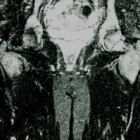
MRI with T2-weighted coronal scans with fat suppression technique shows two areas of bone marrow edema near the growth plate (arrows).
Pelvis AP x-ray performed in the frog lateral position shows the pathological angle between the head and the neck of the left femur (B) and the normal aspect of the controlateral femur head (A).

1. Radiological Findings
Based on the pelvic X-ray (AP view) and MRI provided by the patient, the main findings include:
- The left femoral head is significantly displaced downwards. The angle between the femoral neck and femoral head is abnormal (“slippage” sign), consistent with the typical radiographic features of Slipped Capital Femoral Epiphysis (SCFE).
- Klein’s line (a tangential line along the lateral border of the femoral neck) does not normally intersect the lateral portion of the femoral epiphysis, suggesting slippage.
- MRI shows signal changes in the subchondral region of the left femoral head and neck, with evidence of bone marrow edema; there may be mild joint effusion.
- The joint space is still acceptable, and there is no obvious large-scale involvement of surrounding soft tissue or significant soft tissue swelling.
2. Potential Diagnoses
Considering the patient’s age (13-year-old male), symptoms (left hip pain both during activity and at rest), and imaging findings, possible diagnoses include:
- Slipped Capital Femoral Epiphysis (SCFE): A common hip pathology in adolescents characterized by posterior and inferior displacement of the femoral head relative to the femoral neck. Radiographic signs include an abnormal Klein’s line and posterior-inferior displacement of the femoral head.
- Other possibilities:
- Femoral head necrosis (AVN): Primarily seen in cases of compromised blood supply or may coexist with SCFE, but radiologically often shows collapse or distinct ischemic areas, differing from the MRI findings here.
- Transient synovitis or other inflammatory conditions: Typically presents with acute onset of pain, less likely to show persistent pain or clear structural displacement.
- Congenital hip deformities (e.g., developmental dysplasia): Usually presents earlier in life; a unilateral pronounced slip is less common.
Given the marked evidence of slippage on imaging, SCFE remains the most likely diagnosis.
3. Final Diagnosis
Combining:
- The patient’s age and sex (most common in males between 9 and 15 years old).
- Clinical symptoms (hip pain, restricted range of motion).
- Imaging findings (femoral head displacement on X-ray, abnormal Klein’s line, bone marrow edema on MRI).
The most likely diagnosis is: Left Slipped Capital Femoral Epiphysis (SCFE).
4. Treatment and Rehabilitation Plan
Recommendations for the treatment and rehabilitation of Slipped Capital Femoral Epiphysis include:
-
Treatment Strategy:
- In cases of acute slippage, closed reduction under anesthesia may be performed, followed by internal fixation with screws or a plate to stabilize the femoral head and prevent further slippage.
- If the slip exceeds half the diameter of the femoral head, an osteotomy of the femoral neck or the intertrochanteric region may be required to restore normal hip biomechanics.
- If the contralateral hip is at high risk for slippage or shows mild signs of slip, prophylactic internal fixation may be considered.
-
Rehabilitation and Exercise Prescription:
- Early Phase (0–6 weeks post-op or post-fixation):
- Use of crutches with minimal weight-bearing; avoid heavy weight-bearing and strenuous activities.
- Under professional guidance, perform isometric muscle contractions of the lower limbs to enhance circulation and prevent muscle atrophy.
- Middle Phase (6–12 weeks post-op):
- As bone healing progresses, gradually introduce partial weight-bearing with the assistance of crutches.
- Begin gentle range-of-motion exercises for the hip, such as active or passive flexion and rotation exercises, while avoiding excessive adduction and external rotation.
- Late Phase (12 weeks post-op and beyond):
- Once the hip is stable, gradually introduce resistance exercises for the lower limbs (e.g., using resistance bands) and light strength training.
- Target the flexors and extensors of the hip with specific strength exercises; progressively transition to jogging, swimming, and other low-impact aerobic activities.
- FITT-VP Principle:
- Frequency: Rehab training 3–5 times per week, adjusted according to individual recovery.
- Intensity: Should not provoke significant pain or discomfort; increase gradually.
- Time: 20–30 minutes per session, or gradually extended depending on the post-op phase.
- Type: Focus on joint mobility and strength exercises, transitioning to aerobic exercise over time.
- Progression: Increase loading and range of motion as hip stability improves; avoid abrupt or excessive training.
- Safety Precautions:
- Avoid direct external impact on the hip post-operatively. Follow medical advice for regular check-ups to monitor for re-slippage or avascular necrosis.
- If there is significant pain, swelling, or a marked decrease in the range of motion, seek a professional evaluation promptly.
- Early Phase (0–6 weeks post-op or post-fixation):
Disclaimer: This report is a reference analysis based on existing examination findings and medical literature. It cannot replace in-person consultation or professional medical advice. The specific treatment plan should be determined by a professional orthopedic or sports medicine physician in accordance with the patient’s actual condition.
Human Doctor Final Diagnosis
Left femoral epiphysiolysis