Osteoblastoma of the femur


Clinical History
Female patient with chronic left hip pain undergoing an X-RAY, CT, bone scan and MRI examinations of the left femur.
Imaging Findings
The patient with a four- year history of mild local pain in the left hip, who did not respond to medical treatment at other clinical divisions, has been referred to our clinic. Slight restriction of motion was found on physical examination. She had no history of trauma or any systemic disease and laboratory test results were normal. Plain X-RAY, CT, MRI and bone scan were performed followed by an excisional biopsy. Pathology reports were in accordance with the osteoblastoma.
Discussion
Osteoblastoma is a rare, benign, primary tumor of bone composed of a richly vascular connective tissue stroma in which there is active production of osteoid and primitive woven bone (3). Accounting for approximately 1% of all primary bone tumors and 3% of all benign bone tumors, osteoblastoma, is usually diagnosed in the second and third decades of life, with the peak range being the second decade. A 2:1 male predominance has been noted (2,3). Most common locations for osteoblastoma, in order of frequency, are the spine, femur, foot and ankle (3). Within the spine, the neural arch is primarily affected. Also, multicentric osteoblastomas have rarely been reported (2,3). Pain, scoliosis, local muscle spasm, rigidity and neurologic deficits are clinical manifestations of osteoblastoma (3,4). Osteoblastomas may be medullary, cortical or subperiosteal in location (4).The variation in the radiologic appearance of osteoblastoma is due to the anatomic location of the lesion.Radiographs usually show an expansile, well-circumscribed, lytic lesion with matrix ossification and mild surrounding sclerosis (1,3,4). Osteoblastoma tends to lead to cortical expansion in 75-94%, and cortical destruction in 20-22%. Other imaging methods such as bone scintigraphy, CT and MRI may provide information about the extent of the lesion (4). Osteoblastoma has low to intermediate signal intensity on T1-weighted MR images and high signal intensity on T2 weighted images.Edema in the surrounding soft tissues and in the bone marrow beyond the tumor margins is prevalent.We can see foci of signal void corresponding to calcifications on both T1 and T2 weighted images. After the administration of a gadolinium compound, tumor and surrounding inflammatory reaction enhance, which may simulate malignant process as a result of extensive inflammatory response (1,4). An osteoid osteoma, a bone abcess, an aneurysmal bone cyst, a chondromyxoid fibroma, an enchondroma, and an osteosarcoma should be seen for the differential radiologic diagnosis of osteoblastoma (1,2,4). Although osteoblastoma and osteoid osteoma share a common age range and similar microscopic appearance, both being osteoid-producing lesions, they differ clinically and radiologically. The size of the nidus is the most important criterion to differentiate between osteoblastomas and osteoid osteomas. By definition, the maximum diameter of the nidus is 2 cm in osteoid osteoma.Larger lesions are categorized as osteoblastomas due to the absence of limited growth potential, which is a feature of osteoid osteoma. Lack of characteristic pain in osteoblastoma is another distinctive feature that can be helpful (1,2,3).The main complaint is dull, localized pain of insidious onset, which is less severe than that of osteoid osteoma.It is not nocturnal and respond to salicylates in 7% of the cases (3,4). In osteoid osteoma pain is more severe at night but is dramatically relieved by salicylates within approximately 20 to 25 minutes .This typical history serves as an important clue to the diagnosis. Their natural histories also differ; that is, whereas osteoid osteoma tends toward regression, osteoblastoma tends toward progression and even malignant transformation (2).Marginal en bloc excision, curettage with bone graft and internal fixation, radiation and chemotherapy are the choices of treatment in osteoblastoma (2,3,4).
Differential Diagnosis List
Final Diagnosis
Osteoblastoma of the femur
Liscense
Figures
Figure 2: CT of the left proximal femur

Figure 3: Bone scintigraphy

Figure 4: Axial T1WI MR

Figure 1: X-ray of the left femur
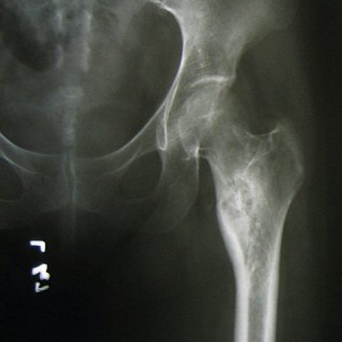
Figure 5: Axial fat-supressed T2WI MR

Figure 6a & b: A coronal STIR


Figure 7: Axial contrast-enhanced T1WI MR

Figure 8: Coronal fat-supressed contrast-enhanced T1WI MR

Figure 9: Microscopic examination

1. Radiological Findings
Based on the provided X-ray, CT, bone scan, and MRI images, the following can be observed:
• The lesion is located in the proximal left femur (left hip region), appearing as eccentric, expansile bone destruction with relatively well-defined margins.
• CT shows partial debris-like or patchy calcifications within the lesion, with focal cortical expansion and thinning, and sclerotic areas in some regions.
• On MRI, T1-weighted images mainly show low to intermediate signal intensity, while T2-weighted and fat-suppressed sequences show high signal intensity. Significant edema is noted in the surrounding and adjacent soft tissues.
• After contrast enhancement, both the lesion and surrounding reactive areas exhibit significant enhancement, with notable inflammatory reaction in the local soft tissue.
• The bone scan demonstrates localized radiotracer uptake, consistent with an active bone lesion.
2. Possible Diagnoses
- Osteoblastoma: Commonly seen in young patients, frequently occurring in the posterior elements of the spine and long bones. The typical presentation includes expansile bone destruction, partial calcification within the lesion, and marked enhancement, correlating with the patient’s complaint of persistent dull pain.
- Osteoid Osteoma: Pathologically similar to osteoblastoma, but the typical nidus usually measures less than 2 cm. Pain often worsens at night and quickly relieves after administration of salicylates. In this case, the lesion is significantly larger than 2 cm, and the pain does not display the typical severe nocturnal presentation.
- Chondromyxoid Fibroma: Can present as an eccentric, expansile lesion, usually exhibiting cartilaginous signals with characteristic marginal calcification or a sclerotic rim. The findings in this case are not fully consistent with its typical presentation.
- Osteosarcoma: Frequently seen in adolescents. It often shows aggressive borders, irregular bone destruction, and periosteal reactions such as “sunburst” or Codman’s triangle. Overall, this case appears more benign, but pathological confirmation is still required.
3. Final Diagnosis
Considering the patient’s age (29 years), chronic left hip pain, radiological findings (expansile bone destruction, partial internal calcifications, a highly enhancing lesion with relatively well-defined margins, and typical histological features), as well as pathological results, the most likely diagnosis is “Osteoblastoma.”
For further confirmation, a complete pathological examination can be performed after surgical resection or curettage of the lesion.
4. Treatment Plan and Rehabilitation Program
Treatment Strategy:
1) Given the characteristics of osteoblastoma, the first choice is surgical intervention, such as curettage or marginal en bloc excision. Bone grafting or internal fixation may be considered if necessary to maintain skeletal stability.
2) For cauterization of the lesion or areas not amenable to complete resection, radiotherapy or other adjuvant therapies may be considered, but should be carefully evaluated.
3) Postoperatively, regular follow-up visits and imaging reviews are recommended to monitor for recurrence or malignant transformation.
Rehabilitation and Exercise Prescription (FITT-VP Principle):
• Early Stage (from postoperative to about 6 weeks): Focus primarily on reduced or partial weight-bearing, along with joint range of motion and muscle strength exercises.
◦ Frequency: 3–5 times per week, with rest days in between.
◦ Intensity: Aimed at alleviating pain and improving joint mobility, avoiding severe pain.
◦ Time: 15–20 minutes per session, performed in sets.
◦ Type: Seated or supine exercises for joint flexion and extension, straight-leg raises, and isometric contractions of the hip musculature.
◦ Progression: If local stability and healing permit, gradually increase the range of motion and weight-bearing.
• Intermediate Stage (6–12 weeks): Gradually increase weight-bearing and joint function training.
◦ Frequency: 3–5 times per week.
◦ Intensity: Increased progressively according to clinical healing, closely monitoring pain and joint function.
◦ Time: 20–30 minutes per session, which can be extended gradually.
◦ Type: Partial weight-bearing walking training, seated/standing muscle strengthening with resistance bands or light resistance equipment, ensuring proper joint alignment.
◦ Progression: Transition to full weight-bearing as tolerated, using crutches or walkers for support as needed.
• Late Stage (12 weeks and onward): Emphasize comprehensive recovery of muscle strength and joint coordination.
◦ Frequency: 3–5 times per week.
◦ Intensity: Moderate to moderately high intensity, depending on bone healing status.
◦ Time: Can be extended to 30–45 minutes per session.
◦ Type: Jogging, cycling, or swimming for aerobic exercise, combined with hip-strengthening exercises. Proprioception and stability training can be added if necessary.
◦ Progression: Adjust exercise load based on follow-up evaluations and individual recovery, avoiding overtraining and stress injuries.
Disclaimer
This report is based on the available information for preliminary reference and does not replace an in-person consultation or professional doctor’s guidance. Specific treatment plans should be determined by the patient’s actual condition, surgical outcomes, and comprehensive evaluation by a qualified physician.
Human Doctor Final Diagnosis
Osteoblastoma of the femur