半肢性骺发育不良(Trevor病)


临床病史
一名6岁男孩因右腕畸形来院就诊。
影像学表现
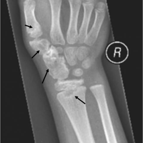
平片检查显示,右手腕存在不规则骨化病变,与桡骨远端骺、舟骨、大多角骨以及第一掌骨近端骺相连续(参见图1)。此外,还可见舟骨和大多角骨整体增大。影像学特征提示半侧骺发育不良(Trevor’s病),累及腕骨以及桡骨和第一掌骨的骺,即典型的Trevor’s病形式。
磁共振(MR)检查证实,这些骨质突起与相应骨在皮质和骨髓层面相连续,并且表现出骨性和软骨性成分,因此呈现出类似骨软骨瘤的外观(参见图2)。腕部肿块与关节积液的共同存在导致机械负担增大,从而引发相关症状。
典型的影像学(包括X线和MR)表现以及患者所处的年龄段对于诊断这一不常见部位的典型型半侧骺发育不良至关重要,及时的诊断与转诊有助于避免出现早期关节退变。
病情讨论
Dysplasia epiphysealis hemimelica (DEH)是一种罕见的骨发育性异常,其特点是在骨骺、骺骨突起、跗骨、腕骨或偶尔在籽骨处出现异常且不对称的骨软骨增生,这些增生在组织学上与普通骨软骨瘤并无区别。历史上,DEH曾有多种名称(tarsomegaly、tarsoepiphyseal aclasis、Trevor-Fairbank病),以体现其临床多样性。
据报道,DEH的患病率约为百万分之一。其病因尚不明确,目前尚未发现任何遗传模式,可能与胚胎期异常相关。
DEH的常见发病年龄为2-14岁,男孩的发病率是女孩的3倍。初始症状包括单侧、无痛、不对称、坚硬的关节肿胀以及活动范围受限;之后可能出现疼痛、肢体萎缩、关节外翻或内翻畸形,有时会出现肢体长度差异。
这种骨软骨增生通常呈半边性,更常见于下肢内侧(远端股骨>近端胫骨>距骨>跗舟骨>第一楔骨),极少数情况可累及上肢及籽骨。
根据Azouz的分类,DEH可以分为局限型(仅累及单块骨)、典型型(约占病例的2/3)或广泛型。典型型表现为同一肢体中有多块骨骼受累,如本例所示。广泛型或严重型则累及整个肢体。
通常依赖影像学(X线)诊断。典型的X线表现为不对称的软骨化中心伴有骨性突出,并可见散在的点状钙化,可能融合成分叶状不规则肿块,随后骨化并与其下方骨骼连接。还可见干骺端继发性变宽并伴管状结构不良。应进行全身骨骼检查以排除其他受累部位。
CT检查有助于证实病变与骨皮质和骨髓的连续性。MR检查可补充X线评估,明确病变的软骨或骨软骨性质及其来源于骨骺,并可更好地显示病变的确切位置和范围、关节受累情况及解剖分界。鉴别诊断包括“牵拉型”外生骨疣及其他类似骨软骨瘤的骨赘;孤立或多发性骨软骨性肿瘤(骨软骨瘤、多发骨软骨瘤、软骨增殖病);以及罕见的慢性婴儿期神经、皮肤和关节(CINCA)综合征。
尽管尚无恶性转化的报告,DEH仍可能导致相当程度的疾病负担。病变可随着骨骼发育而增大,引起骨骺过早闭合、关节畸形及肢体长度差异。关节面不规则可能导致早期继发性骨关节炎,并常伴随疼痛、畸形或机械功能丧失。
建议在患者骨骼成熟之前持续监测,以评估疾病进展。由于DEH位于骨骺区域,更容易出现症状,因此比孤立的骨软骨瘤更常需要手术干预。值得注意的是,DEH可能多年未被识别,从而延误了及时的骨科治疗并导致功能障碍。手术应在早期进行,其目标是改善关节相合性,从而预防或减轻后续出现的继发性骨关节炎并改善预后。即使是非完全切除也可能减轻症状,但畸形复发的报道率较高。
鉴别诊断列表
最终诊断
骨骺半侧发育不良(Trevor病)的经典类型
证书
没有可翻译的内容。
图像分析
右手腕的核磁共振检查


右腕X线片

医学影像分析报告
一、影像学发现
根据患儿右腕部X线与MRI影像所示,可见右腕远端桡骨与腕部数块骨骼(包括腕骨部分)出现不对称、分叶状的骨质增生突起,呈明显外凸状,且与原骨的骨皮质及骨髓呈连续性。病变区可见软骨样信号与散在小片状或团块状钙化影,MRI信号以软骨成分为主,T2WI上呈高信号,骨性成分呈中低信号。腕关节周边关节面轻度不规则,关节间隙可见一定程度受累,但尚未见明显骨折征象或明显软组织损伤。总体表现示意在右腕关节部位出现多个大小不一、边缘不整、向关节面突出的骨软骨性病变。
二、潜在诊断
-
骨软骨瘤(Osteochondroma)或多发性骨软骨瘤:
该病常表现为与骨骼连续的外生性骨赘,外表覆以软骨帽。但常见于干骺端向外突起,且多集中于长骨远近端干骺端,累及关节面的情况较少。
-
牵引性外生骨疣(“traction” exostosis):
主要见于肌腱或韧带附着点的慢性牵拉所致之骨赘。常累及骨突起附近,但通常不会呈现明显的关节面畸形或多发累及。
-
发育性骨软骨异常(如 Trevor-Fairbank 病 / Dysplasia Epiphysealis Hemimelica,DEH):
常发生于骺板(尤其是关节面的半侧),呈不对称增生。该病可出现关节面变形,病灶与骨髓、骨皮质连续,可见软骨成分和不规则钙化影。特点是好发于儿童,男童更易受累。经常累及下肢,也可见于上肢或腕部。
-
罕见综合征(如 CINCA 综合征)导致的骨异常:
但多合并神经、皮肤等系统表现,病变更为广泛。影像学上也可见多发骨赘样病变,但结合临床症状考虑相对少见。
综上所述,最需重点考虑为发育性骨软骨异常(DEH),亦称 Trevor-Fairbank 病,其外生性骨赘呈现对关节面半侧的侵袭或增生,通常与软骨帽相连,并可导致关节变形。
三、最终诊断
结合患儿年龄(6 岁)、性别(男童)、临床表现(右腕畸形)以及影像学特征(腕关节内侧多个不规则骨软骨突起,与骨骼连续,局部关节面不规则),综合判断,最可能的诊断为 “Dysplasia Epiphysealis Hemimelica(DEH,Trevor-Fairbank 病)”。
如需更进一步明确,可行CT三维重建或关节镜检查等,结合骨病理学检查加以确诊。
四、治疗方案与康复计划
-
治疗策略:
- 病情监测:在患儿未达骨骺闭合前,定期进行影像随访,以评估病灶进展和对关节功能的影响。
- 手术干预:当病变引起明显关节功能障碍、疼痛或显著畸形时可考虑行手术切除(部分或完整切除病变骨质),以纠正关节面不平或减轻症状。若畸形导致生长障碍或肢体不平衡时,需由骨科医师进一步评估是否需要矫形手术。
- 保守管理:对轻度症状或无功能障碍者,可在骨科和康复科随访下,以保守方式(观察、物理治疗、护具支持等)尽可能维持关节活动度。
-
康复/运动处方建议:
基于患儿右腕关节的畸形及骨质改变,应采用循序渐进、个体化的康复训练方案,遵循 FITT-VP 原则(频度Frequency、强度Intensity、时间Time、类型Type、进阶Progression、体积Volume):
-
早期目标:避免过度负荷与关节应力,保证基本关节活动度和肌力。
- 频度:每周3~4次,间隔1天休息。
- 强度:低强度,主要以恢复关节活动度为主。
- 时间:每次15~20分钟,配合休息和放松环节。
- 方式:腕部轻度主动关节活动,如握拳、腕背伸、腕掌曲及轻度旋前旋后等柔韧练习;避免大负重或牵拉动作。
-
中期目标:增强腕部及前臂肌群力量,改善关节稳定性与功能。
- 频度:每周3~5次,也可根据适应情况逐渐增加。
- 强度:从低强度过渡到中等强度,辅以弹力带或轻小哑铃进行轻负荷阻力训练。
- 时间:每次可延长到20~30分钟,并加强关节灵活性训练。
- 方式:在专业康复师指导下进行腕关节抗阻练习(如借助弹力带进行掌曲/背伸、旋前/旋后训练),保证动作规范。
-
后期目标:进一步提高功能,逐渐过渡到日常活动甚至简单运动。
- 频度:每周可保持4~5次,依功能恢复情况调整。
- 强度:可逐步增加负荷,但仍需避免剧烈冲击或高负荷训练。
- 时间:每次可维持30分钟左右,以不出现痛感或不适为度。
- 方式:结合腕部协调能力和耐力训练,尝试如轻度球类活动或生活技能训练,强化腕关节灵活度与功能。
- 个体化注意:若存在明显疼痛、关节肿胀或活动度降低,应减少训练量并尽快复诊。若有下肢畸形或其他系统损伤,应综合评估全身情况,以保证安全。
-
早期目标:避免过度负荷与关节应力,保证基本关节活动度和肌力。
免责声明
本报告为基于现有影像与临床信息所做的参考性医学分析,不能替代线下面诊及专业医生的诊断与治疗建议;具体诊疗方案应由骨科及相关专科医生综合评估后决定。如病情有任何变化或出现新的症状,应及时就医并获得进一步专业指导。
人类医生最终诊断
骨骺半侧发育不良(Trevor病)的经典类型